
גידולי בלוטת התריס
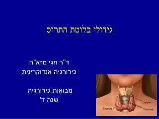
גידולי בלוטת התריס. ד"ר חגי מזא" ה כירורגיה אנדוקרינית מבואות כירורגיה שנה ד'. Indications for Surgery Benign. Benign: Compression symptoms: Dyspnea Dysphagia Hoarseness Hyperthyroidism: Toxic nodule Toxic MNG Graves’ disease Aesthetic concerns. Indications for Surgery Malignant.

גידולי בלוטת התריס
E N D
Presentation Transcript
גידולי בלוטת התריס ד"ר חגי מזא"ה כירורגיה אנדוקרינית מבואות כירורגיה שנה ד'
Indications for SurgeryBenign • Benign: • Compression symptoms: • Dyspnea • Dysphagia • Hoarseness • Hyperthyroidism: • Toxic nodule • Toxic MNG • Graves’ disease • Aesthetic concerns
Indications for SurgeryMalignant • Papillary thyroid cancer(PTC) • Follicular thyroid cancer (FTC) • Medullary thyroid cancer (MTC) • Anaplastic thyroid cancer (ATC) • Lymphoma • Mets
Thyroid Cancer • Most common malignancy of the endocrine system • Increasing incidence • 2% of all new cancer diagnoses • Over 44,000/y expected to be diagnosed in the US
Thyroid nodules • Very common • 5% have palpable thyroid nodules • ~50% have thyroid nodules on US • Only 5% malignant
Case 1 • 24 YOF • Upon shaving identified a 3 cm mass in her front neck
Case 1 • Physician – • History • Thyroid function (hyper/hypo) • Risk factors – family history, radiation history • Physical examination • Nodule • Other thyroid nodules • Cervical lymph nodes • Labs • Thyroid function tests
Case 1 • Radiology – Ultrasound: • Most accurate imaging for thyroid nodules • Nodule location • Nodule size (3 dimensions) • Nodule growth • Nodule features: • Microcalcifications • Solid • Lack of Halo / hypoechoic rim • Taller more than wide • Irregular margins • Hypervascular • Hypoechoic • Local invasion
Case 1 • US guided FNAB • Limited to cells (no vascular or capsular invasion) • Equivocal diagnosis
Case 1 • FNAB result – Benign • Management: • Observation • Repeat US • Repeat FNAB (?)
Case 2 • FNAB result – Malignant • Well-differentiated thyroid cancer • Well-differentiated thyroid cancer: • Papillary (PTC) or Follicular (FTC) • Most common (>90% of thyroid cancers) • Very good prognosis (>95% 10YS)
Well-differentiated thyroid cancer Staging ≥ 45 y: • Stage 1 – T< 2cm • Stage 2 – T 2-4cm • Stage 3 – T>4cm, N1a • Stage 4 – M1, N1b < 45 y: • Stage 1 – any T, any N • Stage 2 – M1
Case 2Well-differentiated thyroid cancer • Management: • Depends on size and LN status • ≤ 1 cm – Lobectomy • > 1 cm – total thyroidectomy • Consider prophylactic CLND
Case 2Well-differentiated thyroid cancer • Lateral LND – • FNAB proven involved LN
Case 2Well-differentiated thyroid cancer • Adjuvant therapy: • Selective RAI • TSH suppression • Follow up: • P/E, Tg, Neck US • Up to 30% will require redo surgery (cervical lymph nodes)
Case 3Medullary thyroid cancer • 3-5% of all thyroid cancers • Parafollicular C cells • 75% sporadic • 25% hereditary • MEN IIA (MTC, pheochromococytoma, primary hyperparathyroidism) • MEN IIB (MTC, pheochromocytoma, neurogangliomas) • Familial MTC (non-MEN II)
Case 3Medullary thyroid cancer • Physician – • History • Thyroid function (hyper/hypo) • Risk factors – family history, radiation history • Other endocrinopathies • Physical examination • Nodule • Other thyroid nodules • Cervical lymph nodes • Labs • Thyroid function tests • Calcitonin / CEA • Genetic counseling
Case 3Medullary thyroid cancer • Radiology • Ultrasound – neck • CT – chest abdomen for mets • Management – aggressive!!! • Total thyroidectomy • Central lymph node dissection • Selective lateral lymph node dissection • Tumor size, preoperative US, calcitonin level
Case 3Medullary thyroid cancer • Adjuvant therapy – • No RAI, No TSH suppression • Clinical trials drugs • Follow up – • CEA, calcitonin • Neck US • Prognosis • 75-85% overall 10YS
Case 3Medullary thyroid cancer • Prophylactic surgery • Mutation based: • Level 3 (Highest risk, 883, 918, 922) – • Within age 6-12 months • Level 2 (Higher risk, 611, 618, 620, 634) – • By the age of 5y • Level 3 (High risk, 609, 630, 768, 790, 791, 804, 891) – • By the age of 10y
Case 4Other thyroid cancers • Anaplastic thyroid cancer: • 1% of thyroid cancers • Undifferentiated thyroid cancer • Usually not resectable • Very poor prognosis (5% 5YS) • Thyroid Lymphoma • 1-2% of thyroid cancers • No surgical treatment • CHOP / radiation
Case 5Follicular lesion • Follicular lesion / neoplasm • 15-30% malignancy • Surgeon • Actually has to talk to the patient!! • Options – Lobectomy / total thyroidectomy • Lobectomy – • decreased complications • may not require thyroid replacement • may need ANOTHER surgery if malignant on pathology
ThyroidectomyComplications • Immediate / early • Bleeding • 1-2%, mostly no intervention required • Hematoma requiring urgent drainage – rare • Transient hypocalcemia • Only following total thyroidectomy • 10-20% • Transient hoarseness • 10-20%
ThyroidectomyComplications • Long term • Permanent hypocalcemia • 2-4% • Permanent hoarseness • 1-2% • Permanent hormone replacement therapy • (following thyroidectomy)