Antenatal Care
Antenatal Care . DR. Yasir Katib MBBS, FRCSC Perinatologest. Antenatal care (ANC) goals and strategy. Explain the components and objectives of prenatal goals Describe the frequency and aim of each prenatal visit Genetic counseling and its available tools

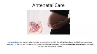
Antenatal Care
E N D
Presentation Transcript
Antenatal Care DR. Yasir Katib MBBS, FRCSC Perinatologest
Antenatal care (ANC) goals and strategy • Explain the components and objectives of prenatal goals • Describe the frequency and aim of each prenatal visit • Genetic counseling and its available tools • Risks and benefits of the genetic tests
ANC Objectives To ensure the birth of a healthy baby with minimal risk for the mother by • Early, accurate estimation of GA • Identification of the patient at risk for complications • Ongoing evaluation of the health status of both mother and fetus • Anticipation of problems and intervention, if possible, to prevent or minimize morbidity • Patient education and communication
ANC • Prenatal care is not a single intervention • “Quantity" Vs. “Quality" of ANC • A systematic review of observational studies and randomized trials concluded that there was no conclusive evidence that prenatal care improved birth outcomes • Randomized trials have also shown that enhanced prenatal care did not result in improved outcomes compared to routine prenatal care
ANC Components • HISTORY • PHYSICAL EXAMINATION • LABORATORY TESTS • DIAGNOSTIC IMIGING • PATIENT EDUCATION • MEDICATIONS
HISTORY • Personal and demographic information • Current pregnancy history (dating) • Past obstetrical history (time, place, GA, complications, sex, weight& currant status) • Personal and family medical history • Past surgical history • Genetic history • Menstrual and gynecological history • Psychosocial information
Physical examination A complete physical examination Specially… • Uterine size and shape • Evaluation of the adenexea • Baseline blood pressure, weight, and height
LABORATORY TESTS1st visit • A standard panel of laboratory tests (Routine) • CBC • Blood gp & antibodies screen • HbS Ag • Rubella • VDRL • Urine C&S • Cervical PAP smear
LABORATORY TESTS1st visit • In high risk population • Chlamydia swab • HIV • TFT
LABORATORY TESTSothers • N. gonorrhea • TB • Toxoplasmosis • HCV • BV • Others HB electrophoresis, cystic fibrosis, phenylalanine level …etc
Follow-up visits • Major goals (PET, malpresentation) Components • Wt, B/P, SPH, FH auscultation, FM, lie, presentation • Patient’s concerns • Education • Risk identifications
Follow-up visits Uncomplicated pregnancies • Every 4 weeks until 28 wks • Every 2 to 3 weeks from 28 to 36 wks • Weekly until delivery
Follow-up visits Laboratory follow-up • GDM screening at 24-28 wks • CBC & antibodies screen • GBS screening (recto-vaginal swab) at 35-37 wks
DIAGNOSTIC IMIGING Ultrasound • Minimum requirement • Usefulness • Limitations Others • MRI
1st Trimester U/S • Confirm pregnancy • Viability • Assess GA (Dating) • Multiple gestations (chorionicity / amnionicity) • Maternal pelvic anomalies AIUM Standards and Guidelines
2nd Trimester U/S Fetal normality: • • Head shape and size and internal structures (cavum pellucidum, cerebellum, ventricular size at atrium < 10 mm) • • Spine: longitudinal and transverse • • Abdominal shape and content at level of stomach • • Abdominal shape and content at level of kidneys and umbilicus • • Renal pelvis < 5 mm anterior–posterior measurement • • Longitudinal axis abdominal–thoracic appearance (diaphragm and bladder) • • Thorax at level of a four-chamber cardiac view • • Arms: three bones and hand (not counting fingers) • • Legs: three bones and foot (not counting toes)
VALUE OF PRENATAL GENETIC DIAGNOSIS:Why do we do it? • Reassurance • Increases options • Antenatal fetal treatment • Preparation for outcome • Avoidance of obstetric complications • Selective termination
Maternal age-based screening Rationale 70% 30% 1/200 risk of miscarriage 1/200 chance of abnormality = =
10 1 0.1 0.01 0.001 0.0001 20 25 30 35 40 44 Years Assessment of Background Risk Maternal Age • Trisomy 21 47xxx/xxy/xyy Risk % • Trisomy 18 • Trisomy 13 45x Triploidy Courtesy Dr J Johnson and the Fetal Medicine Foundation
100 47xxx/xxy/xyy 80 60 Trisomy 21 40 45x 20 Trisomy 18 Trisomy 13 0 Triploidy 10 14 18 25 30 35 40 Weeks Assessment of Backround Risk Gestational Age % Courtesy Dr J Johnson and the Fetal Medicine Foundation Snijders et al 1999
Screening at 11-20 wks 10 12 14 16 18 20 wks • Allows adjustment of age-related risk • Improved detection rate Courtesy Dr J Johnson and the Fetal Medicine Foundation
A-Non-invasive prenatal diagnostic tests • Nuchal Screen • Nuchal plus biochemistry • Maternal Serum Screen • Ultrasound
Nuchal Translucency 1- “the skin is deficient in elasticity. . . . . . too large for the body” Langdon Down Observations on an ethnic classification of idiots. Clinical Lecture Reports, London Hospital 1866;3:259. Courtesy Dr J Johnson and the Fetal Medicine Foundation
Nuchal Translucency • Gestation 11-14 wks • CRL 45-84 mm • Mid-sagittal view • Image size >75% • Neutral position • Away from amnion • Maximum lucency • Calipers on-to-on Courtesy Dr J Johnson and the Fetal Medicine Foundation
Significance of increased nuchal fluid • Chromosome abnormalities • Birth defects (cardiac, d.hernia) • Genetic syndromes • Increased mortality (> 3.5 mm) Courtesy Dr J Johnson and the Fetal Medicine Foundation
Pathophysiology of increased nuchal translucency • Abnormal or delayed lymphatic development • Venous congestion • Cardiac failure • Altered composition of extracellular matrix • Failure of lymphatic drainage due to fetal hypokinesia • Fetal anemia or hypoproteinemia • Congenital infection • Courtesy Dr J Johnson and the Fetal Medicine Foundation
First Trimester Biochemical Markers 2- • PAPP-A: Pregnancy associated plasma protein A • Produced by placental trophoblast • Increases in 10-14 week period • Lower in DS pregnancies (0.43 MOM) • Associated with 42 % DR at 5% FPR. • Free - hCg: Free subunit of human chorionic gonadotrophin. • Placental protein • Decreases in T1 like total hGC • Higher in DS pregnancies (1.79 MOM) • Associated with 23% DR at 5% FPR • Courtesy Dr J Johnson and the Fetal Medicine Foundation
Detection Rate 89% 100 72% 80 60% % 60 30% 40 20 0 Age ßhCG PAPP-A Age Age NT All ßhCG PAPP-A Screening at 11-14 wks Fetal NT + Maternal age + ßhCG + PAPP-A at 11-14 wks Invasive Testing 5% • Courtesy Dr J Johnson and the Fetal Medicine Foundation
3- Maternal Serum Screen • Three biochemical markers • BHCG • AFP • Estriol • Gives age adjusted risk • Screens for Down’s and NTD • 15-20 wks
MSS 0 1/1 1/378 Risk = age X OR BHCG X OR uE3 X OR AFP
MSS Limitations • Sensitivity 70 % • Specificity 95% ( False positive 5%) Two reasons for a false positive: • Wrong dates • Twins
4- Ultrasound • Ultrasound screening(detailed scan) • 18-20 weeks
B- INVASIVE TECHNIQUES FOR EARLY PRENATAL TESTING • Chorionic Villus Sampling • Amniocentesis
Amniocentisis http://wchs.health.wa.gov.au/health/a/amnio.htm
Amniocentisis • Performed at or more 15 weeks • Takes 2-3 weeks for Karyotype • Pregnancy loss risk 1/200 • Can get result of trisomies 13,18,21 and Turners Syndrome X0 in 48 hours with FISH (Florescent Insitu Hybridization)
Chorionic Villus Sampling • Performed at 10-14 weeks • Takes 2-3 weeks for the result • Pregnancy loss rate 1/100 • Operator experience important • Link to limb abnormalities (still controversial) • Placental mosaicism up to 3%, but little effect on outcome
Post Test • A standard panel of laboratory tests (Routine) dose not include • Rubella • TFT • VDRL • Urine C&S • Cervical PAP smear
Post Test First trimister scan does not include • Fetal morphology • Viability • Assess GA (Dating) • Multiple gestations (chorionicity / amnionicity) • Maternal pelvic anomalies
Post Test Which of the following is not a cause for an abnormal MSS • Down’s syndrome • IUFD • Twins • Wrong dates • Congenital heart disease
Post Test A false positive on the MSS occurs in 1/20 tests • True • False
Post Test List Two advantages of Amniocentesis over CVS • Decreased miscarriage rate • Lower risk of mosaicism • No association with limb reductions
Post test Which of the following tests is the best at detecting Down’s Syndrome • MSS • Nuchal • Nuchal plus PAPP A and Free BHCG
Post Test What is the miscarriage rate with amniocentesis? • 0.5% • 3% • 5% • 10%
Post Test List one advantage of CVS over amniocentesis • Early results