Download

1 / 39
530 likes | 3.3k Vues
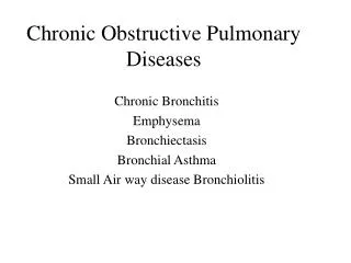
Chronic Obstructive Sialadenitis & Sialendoscopy. Anatomy--Salivary gland . Parotid Ductal System Nicolaus Stenonius 1660. Stensen’s duct. Anatomy--Salivary gland. Bartholin’s duct. Submandibular Ductal System Thomas Wharton, 1659 Sublingual Ductal System Casparus Bartholinus, 1690.

E N D
Anatomy--Salivary gland Parotid Ductal System Nicolaus Stenonius 1660 Stensen’s duct
Anatomy--Salivary gland Bartholin’s duct • Submandibular Ductal SystemThomas Wharton, 1659 • Sublingual Ductal SystemCasparus Bartholinus, 1690 Wharton’s duct
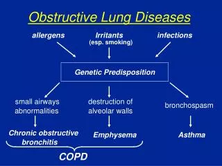
Chronic Obstructive Sialadenitis • Chronic Obstructive Parotitis • Submandibular sialadenitis
Chronic Obstructive Parotitis • Etiology • Clinical manifestations • Diagnosis • Treatment
Etiology ofChronic Obstructive Parotitis • Scar • Sialolithiasis • Anatomy
Clinical manifestations • Recurrent swelling and pain of the gland • Purulent discharge
Diagnostic methods • Plain radiographs • Sialography • Ultrasound • Scintigraphy(闪烁扫描法) • CT • MRI
Sialography “Sausage like” appearance of enlarged duct
MRI Appearance of enlarged duct
Sialography Endoscopy Sialendoscopy Dilation Stenosis
Differentiating diagnosis • Chronic recurrent parotitis • SjÖgren syndrome
Treatment • systemic antibiotic administration • Sialogogues(促唾剂) • Gland massage • Drug lavage(灌洗) and perfusion • Duct ligation • Parotidectomy • Tympanic(鼓室) neurectomy
Etiology of Submandibular sialadenitis • Sialolithiasis • Trauma • Infection • Foreign body
1. Anatomy Upwarding route Longer duct Curve duct 2.Components of saliva Mucus protein Calcium content SialolithiasisReasons of arising
Manifestations • Intermittent swelling of the gland • Aggravating with taking food • Acute infection
Diagnostic methods • plain radiographs • sialography • ultrasound • CT scan • Sialoendoscopy
Plain radiographs One Two Three
Sialography & CT Sialolith
Sialoendoscopy Stone in second branch duct Stone embedded Stone in main duct
Differentiating diagnosis • Tumor in sublingual gland • Tumor in submandibular gland • KÜtter tumor • Space infection in submandibular region • Lymphadenopathy
Traditional treatment • Intraoral route • Sialadenectomy via external approach
New technique Sialendoscopy • Diagnostic Sialendoscopy • Interventional Sialendoscopy
History of the Sialendoscopy • 1991 KatzFlexible mini-endoscope • 1993 KonigsbergerEndoscopic intracorporeal lithotripsy • 1994 Arzoz Endoscopic intracorporeal lithotripsy Nahlieli Sialendoscopy • 1995 Marchal Sialendoscopy • 1999 Our Dept. Yu
Diagnostic Sialendoscopy First generation branches Second generation branches
Types of obstructions • Sialolith • Polyps • Stricture • Kink • Foreign body • Anatomic malformation
Sialolith 80%~90% in Submandibular gland First branch of duct Main duct
Polyps and Mucous Plug Sialolith 10%~30% in Parotid gland Mucous Plug Sialolith?
Interventional Sialendoscopy • Grasping wire basket • Biopsy forceps • Balloon-tipped catheter • Custom papilla dilator • Electrohydraulic lithotripter • Holmium laser probe
Grasping wire basket diameter<4mm
Balloon-tipped catheter Ductal Stenosis Stenosis in the second branch Close-up view of the same site
Electrohydraulic Shockwave Lithotripsy • Stone was fragmentized by Lithotripter • Debris extracted by wire basket diameter>4mm
Holmium laser probe Laser fragmentation • Stone debris extracted by wire basket
Sialendoscopy in our department 2002 1999 2003