Download
1 / 63
670 likes | 1.28k Vues
Introduction to Cardiovascular Pathology - Fred Clayton. Systemic Pathology of Congestive Heart Failure Pathology of Myocarditis Pathology of Cardiomyopathy Dilated Cardiomyopathy Hypertrophic Cardiomyopathy Restrictive Cardiomyopathy. Congestive Heart Failure.

E N D
Introduction to Cardiovascular Pathology- Fred Clayton • Systemic Pathology of Congestive Heart Failure • Pathology of Myocarditis • Pathology of Cardiomyopathy • Dilated Cardiomyopathy • Hypertrophic Cardiomyopathy • Restrictive Cardiomyopathy
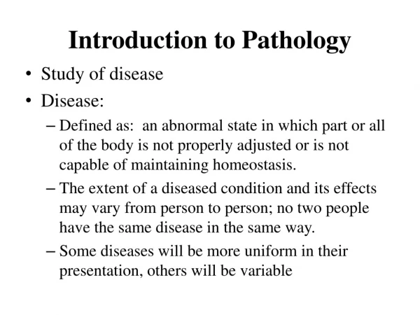
Congestive Heart Failure • Cardiac output insufficient for metabolic requirements of the body • Systolic dysfunction – decreased myocardial contractility • Diastolic dysfunction – insufficient expansion for ventricular volume • Problems are accentuated by increased demand – high output heart failure
CHF – Body’s Compensation • Tachycardia • Frank-Starling – increased End Diastolic Volume • Myocardial hypertrophy • Renin-angiotensin-aldosterone system • Catecholamines – positive inotropic effect • Adrenergic redistribution of blood flow • Increase oxygen extraction from hemoglobin
Left-sided Heart Failure • Ischemic heart disease • Hypertension • Aortic and mitral valve disease • Myocardial disease
Lungs – Pulmonary edema • Dyspnea – breathlessness • Orthopnea – dyspnea lying down • Paroxysmal nocturnal dyspnea – extreme dyspnea
Lung – Pulmonary Edema – pale pink edema fluid filling alveoli
Lung – alveolar hemorrhage, heme-filled macrophages “heart failure cells”, with iron stain to right
Kidneys – reduced perfusion • Ischemic tubular necrosis / ATN • Prerenal azotemia
Brain in CHF – cerebral hypoxia • Irritability • Loss of attention span • Restlessness • Stupor • Coma
Right-sided heart failure • Pure cor pulmonale • Consequence of left-sided failure • Myocardial – myocarditis, cardiomyopathy, constrictive pericarditis
Right failure - systemic effects • Liver – chronic passive congestion • Spleen – congestive splenomegaly • Kidneys – congestion and hypoxia • Sub-Q – peripheral edema and anasarca • Pleural space – effusions • Brain – venous congestion and hypoxia • Portal - ascites
Liver – chronic passive congestion – blood pools near the central veins
Liver – chronic passive congestion – blood pools near the central veins
Liver – chronic passive congestion – red cell pooling near central veins and pericentral necrosis of the hepatocytes
CHF – final pathway to death • Ischemic heart disease • Hypertensive heart disease • Valvular heart disease • Cardiomyopathy • Myocarditis • Specific heart muscle diseases
Myocarditis Etiology • Viral – Coxsackie A, ECHO, Influenza • Chlamydia and Rickettsia – psittaci & typhi • Bacteria – diphtheria, TB, Strep • Fungal & Protozoa – Trypanosomes, Toxo • Hypersensitivity – SLE, RHD, drugs • Physical Agents – Radiation • Idiopathic – Giant cell myocarditis
Myocarditis Morphology • Gross –dilated, flabby heart, pale patches with hemorrhage • Microscopic – interstitial inflammatory infiltrate with myocyte necrosis, fibrosis • Mononuclear cells – idiopathic or viral • Neutrophils – bacterial • Eosinophils –hypersensitivity or protozoa • Granulomatous – TB or sarcoid
Dilated, globoid heart in myocarditis
Myocarditis – meets Dallas criteria of a T lymphocyte infiltrate and myocyte necrosis or dropout. This is usually either viral or of unknown cause.
Diphtheria myocarditis – due to a toxin rather than bacterial invasion. There is some inflammation, myocyte changes (see the big nucleolus). Myocyte necrosis (not shown) also happens.
Giant Cell Myocarditis • Myocyte necrosis • Multinucleated giant cells • Lymphocytes, plasma cells, macrophages, eosinophils, and neutrophils • Often fulminant, rapid progression to death • Differential diagnosis – cardiac sarcoidosis
Dilated Cardiomyopathy • Gross – increased weight, dilatation, endocardial fibrosis, normal valves and coronary arteries • Microscopic – myocyte hypertrophy, myofibrillar loss and interstitial fibrosis • Etiology – viral, genetic, toxins • Clinical significance – heart failure & death
Cardiomyopathy – trichrome stain showing extensive fibrosis (blue) between the myocytes. The myocytes also vary in size, and some have partial loss of myofibrils.
Loss of fibrils in cardiomyopathy. The myocyte at lower left is about normal; the others have an extensive loss of myofibrils.
Cardiomyopathy – loss of fibrils and a small contraction band in the top center.
Hypertrophic Cardiomyopathy • Hypertrophy of ventricular septum (95%) • Disarray of myofibers (100%) • Volume reduction of ventricles (90%) • Endocardial thickening of LV (75%) • Mitral valve leaflet thickening (75%) • Dilated atria (100%) • Abnormal intramural coronaries (50%)
Hypertrophic cardiomyopathy – myofiber dysarray – not all fibers are pulling the same direction. Thus the contraction is ineffective. However, the cardiac conduction system can have these same problems, which might cause the arrhythmias and sudden death these patients tend to die of.
Hypertrophic Cardiomyopathy • Etiology – hereditary, mostly autosomal dominant, can appear sporadically • Clinical significance – syncope, arrhythmias and sudden death with a risk of 2-6% per year • Cannot equate with hypertrophy alone! There is variation in heart size without disease. Large hearts correlate with endurance (Secretariat, Lance Armstrong).
Restrictive Cardiomyopathy • Amyloidosis • Endomyocardial fibrosis – subendocardial fibrosis • Loeffler’s endocarditis – eosinophilic infiltrate • Endocardial fibroelastosis
Amyloidosis – notice the pink material between the myocytes.