STRUCTURE OF CHROMATIN
310 likes | 2.33k Vues
STRUCTURE OF CHROMATIN . Lindsey Suttle, Aaron Alejandro, Christine Nam and Aruna Iyer . BUILDING BLOCKS.

STRUCTURE OF CHROMATIN
E N D
Presentation Transcript
STRUCTURE OF CHROMATIN Lindsey Suttle, Aaron Alejandro, Christine Nam and Aruna Iyer
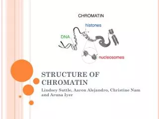
BUILDING BLOCKS • Nucleosome: The basic, beadlike unit of DNA packaging in eukaryotes, consisting of a segment of DNA wound twice around a protein core composed of two copies of each of four types of histone. • Histone: A small protein with a high proportion of positively charged amino acids that binds to the negatively charged DNA • It plays a key role in its chromatin structure.
PROTEIN SCAFFOLDING • Protein Scaffolding: The H1 histone that attaches to the histones and chromatin for support • Proteins called histones have a high proportion of positively charged amino acids and bind to negatively charged DNA. The DNA-histone complex is chromatin in its most basic structure. Histones are similar in most eukaryotes. Unfolded chromosomes look like beads on a string. Each bead and its DNA is called a nucleosome. The nucleosome bead is DNA wound around a protein core made of two of these histones: H2A, H2B, H3, and H4. H1, another histone attaches to the DNA near the bead when the chromatin undergoes the next level of packing.
CHROMATIN FIBER • Chromatin fiber: the folded complex of DNA and histone proteins that is roughly 30nm in thickness that are very long and not visible with a light microscope. • The chromatin fibers coil up to form chromosomes • Also known as 30-nm chromatin fiber or 30-nm fiber • Looped domain: the loop formed by chromatin fiber • This attaches to a chromosome scaffold made of nonhistone proteins.
2 TYPES OF INTERPAHSE CHROMATIN • Heterochromatin: highly condensed state of chromatin • Visible through a light microscope • Does not undergo transcription • Euchromatin: lightly compacted chromatin • Undergoes transcriptions • Found in both eukaryotic and prokaryotic cells
MODIFICATIONS • DNA methylation: the attachment of methyl groups (-CH3) to DNA bases (after DNA is synthesized) • Methylation can turnoff genes • Demethylation is when the extra methyl groups are removed • Demethylation can activate genes • Protects and stabilizes DNA • Histone acetylation: when an acetyl group (-COCH3) is attached to certain amino acids of histones. • When a histone is acetylated it changes shape making the DNA fit less tightly allowing for other proteins to bind for transcription. • Deacetylation is the removal of acetyl groups.
YOUTUBE • http://www.youtube.com/watch?v=gbSIBhFwQ4s&feature=related
PREVIOUS VOCAB • Nucleic acids • Dehydration synthesis • Light microscope • Proteins • R groups • Mitosis/meiosis
OBJECTIVE TERMINOLOGY • Chromatin • Chromosome • Looped domains • Heterochromatin • Nucleosome “beads” • Histones • Proteins scaffolding • DNA methylation • Histone acetylation • Euchromatin
REVIEW QUESTIONS • What makes up Protein Scaffolding? • Histones, DNA, nucleosomes • Four histones with DNA wrapped twice around the four histones creates what is known as ______ • Nucleosomes • What does DNA methylation do for the DNA? • Turns off genes, protects, and stabilizes DNA • Histone Acetylation occurs to let what other process occur? • Transcription, by making the fit of the DNA looser • What does the looped domain attach to? • The chromosome scaffold (non histone proteins)
REVIEW QUESTIONS CONTINUED • What is the main difference between euchromatin and heterochromatin? • The tightness in which they are compacted. Heterochromatin is highly condensed form of chromatin and euchromatin is lightly compacted • In what type of cells is euchromatin found? • Eukaryotic and prokaryotic cells • Which form of chromatin undergoes transcription? • Euchromatin • Contrast the structure of chromatin before and after Interpahse. • Before Interpahse the chromatin is just chromatin after Interpahse the chromatin is wound around histones and is in the form of chromosomes
JUST FOR FUN • http://www.youtube.com/watch?v=TUFsMY156fc
![[IV] The Role of Chromatin Structure in Control of Gene Expression](https://cdn2.slideserve.com/4473274/slide1-dt.jpg)