Download
1 / 75
780 likes | 1.04k Vues
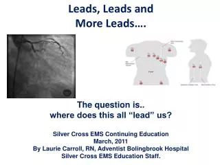
Leads, Leads and More Leads…. The question is.. where does this all “lead” us ? Silver Cross EMS Continuing Education March, 2011 By Laurie Carroll, RN, Adventist Bolingbrook Hospital Silver Cross EMS Education Staff.

E N D
Leads, Leads and More Leads…. The question is.. where does this all “lead” us? Silver Cross EMS Continuing Education March, 2011 By Laurie Carroll, RN, Adventist Bolingbrook Hospital Silver Cross EMS Education Staff.
Welcome to the our first web-based CME offering at Silver Cross EMS! A reminder: this presentation will be recorded for future authorized use. We’d like to begin our first web-based CME offering with a short message from our EMS Medical Director, Dr. Dave. This audio will be heard from your computer speakers, not your phone, so please adjust volume accordingly.
Thank you Dr. Dave! FYI… ILS and BLS providers will not be tested on the 12-lead portion of this presentation, but they will be asked questions about the first half, including cardiac anatomy and other basic life support items.
The Heart • Four chambers • Hollow • Muscular • Dual Circulation • feeds the heart muscle itself • circulates blood outside the heart • lungs for oxygenation and CO2 offload • peripheral supply to tissues, organs and organ systems
Right Heart • Receives unoxygenated blood from • systemic circulation • vena cavae • coronary circulation • coronary sinus • Blood passes from • right atrium • through tricuspid valve • right ventricle • pulmonic valve • pulmonary artery • to lungs
Left Heart • Receives oxygenated blood from pulmonary vein • Blood passes from • left atrium • mitral valve • left ventricle • aortic valve • aorta
Aorta • Oxygenated blood in the aortic root enters the coronary arteries • Most myocardial blood supply occurs during diastole • the aortic valve leaflets are closed and do not obstruct the coronary artery roots • the subendocardial blood vessels are not compressed (as they are during systole) allowing blood to flow into the myocardium itself • in the normal cardiac cycle, diastole is longer than systole
Coronary Artery Disease • Any narrowing of the coronary arteries causes • diminished blood supply • restriction of delivery of electrolytes and nutrients
Who Supplies What? It is often helpful to understand where the supply of blood is coming from, to help us understand which areas of the heart are affected by various strictures/occlusions.
LCA • Left • Left main divides into two branches • Left Anterior Descending (LAD) • Anterior wall of LV • RBB and portions of LBB • Associated with Anterior Wall MI • Circumflex (Cx) • Lateral and Posterior walls of LV • Left atrium • SA node in ~ 30% • Associated with Lateral Wall MI
RCA • Right • Right Atrium (RA) • Right Ventricle (RV) • Inferior and Posterior LV • SA node in ~60% • AV node • Associated with • right ventricular MI • or • dysrhythmias affecting • SA and AV nodes
Area of Injury • Infarct = dead or necrosis • Blockage = causes ischemia
Collateral Circulation • If coronary artery disease and stenosis develop slowly, collateral circulation can develop • When the stenosis is acute, collateral circulation does not have time to develop
Cardiac Rhythm Disturbances • CO (cardiac output) = HR (heart rate) x SV (stroke volume) • Rhythm disturbances can hamper delivery of blood to the myocardium
Normal ECG Review • P wave • Smooth, rounded, upright • P-R Interval • .12 - .20 seconds • QRS • Symmetrical • < .10 seconds • S-T Segment • Isoelectric • T wave • Upright, rounded
Hook ‘em up: • Where do the stickies go, and why? • A lead is a record of electrical activity between two electrodes. Each lead records the average current flow at a specific time in a portion of the heart. • Skin preparation: • dry, hair-free • Placing the electrode. Be sure that the electrode has adequate gel and is not dry.
Trouble shooting EKG Clarity • Equipment Grounded? • Cables attached? • Patient in reclining or semi fowler position? • Patient sitting still? • Skin clean and dry? • Limb leads in place or reversed?
There are three kinds of leads: • Standard Limb Leads • Augmented Leads • Precordial Leads
Standard Limb Leads • (Remember: “lead” may refer to a direction, or placement.) • Lead I: The positive lead is above the left breast or on the left arm and the negative lead is on the right arm.Records the difference of potential between the Left arm and Right arm. • Lead II: The postive lead is on the left abdomen or left thigh and the negative lead is also on the right arm.Records the difference of potential between the left leg and the right arm. • Lead III: The postive lead is also on the left abdomen or left lower lateral leg but the negative lead is on the left arm.Records the difference of potential between the left leg and the right arm.
Augmented Leads • The four limb leads go on the four extremities as follows: • The upper extremities need placement of the electrodes • on the area of the lateral humoral aspect of the arms. • The lower extremities need placement of the electrodes • on the lateral lower legs near the lateral mallelous. • Lead aVR faces the heart from the right shoulder and is oriented to the cavity of the heart. • Lead aVL faces the heart from the left shoulder and is oriented to the Left Ventricle. • Lead aVF face the heart from the left hip and is oriented to the inferior surface of the Left Ventricle.
Precordial Lead Placement • v1 - 4th ICS, R sternal border • v2 - 4th ICS, L sternal border • v3 - midway between v2 & v4 • v4 - 5th ICS, L MCL • v5 - 5th ICS, between v4 & v6 • v6 - 5th ICS, L mid-axillary line Tip Some find it easier to put the leads on in this order v1, v2, v4, v6 Then v3 and v5
You Lookin’ at Me? It is important to look at contiguous leads to determine which area of the heart is affected. Each lead is like a camera lens that “looks” at an area of the heart.
And how do I know if I have the leads in the right place? If everything in Lead I (P, QRS & T wave) is inverted, RA and RL are reversed. Watch for the progression of the R wave in the precordial leads. • “R” WAVE PROGRESSION • The right ventricle depolarizes faster than the left ventricle because it is smaller. • The left ventricle sits to the left and posterior to the right ventricle. • As current spreads leftward through the left ventricle, the height of the R wave in the precordial leads progressively increases. • Normally, in V1 the R wave is more negative and as it progress to V6 the R wave becomes more positively deflected. • “R” wave progression indicates that current is flowing normally through the anterior plane of the heart. (Conover)
ECG Patterns • As contiguous leads look at different parts of the heart, you may see an ischemic pattern that covers a large area or border area between two regions. • For example, if there is ST-segment elevation in leads II/III/aVF/V5/V6, the ischemia appears to be on both the inferior and lateral areas, referred to as inferolateral. • Likewise, there are anterolateral and anteroseptal (like the illustration here) ischemic patterns. • What is an ischemic pattern? The AHA cites “typical ST-segment elevation” as “> 1 mm in 2 or more contiguous leads”
Injury=Elevated ST segment • Signifies an acute process; ST returns to baseline with time • Location of injury can be determined in same • manner as infarct location • Usually associated with reciprocal ST depression • in other leads • If ST elevation is diffuse and unassociated • with Q waves or reciprocal ST depression, • consider pericarditis
Anterior MI • LAD • Most lethal with highest mortality • Can suddenly develop CHB, VF, VT • If present with hemiblocks or BBB, • Can extend to septum and/or lateral walls • Nitrates are desired over fluids
Anterior Infarction • ST elevation without abnormal Q wave • Usually associated with occlusion of the left anterior descending branch of the left coronary artery (LCA)
Lateral or Septal Wall MI Rarely seen alone, usually an extension of anterior or inferior MI Septal – Left Anterior Descending Lateral – Left Circumflex
Pre Intervention Post Intervention
Lateral Infarction • ST elevation with/without abnormal Q wave. • May be a component of a multiple-site infarction • Usually associated with obstruction of the left circumflex artery.
Inferior Wall • RCA • The most common type of MI • Nausea is common • Frequent re-infarction or extends to lateral wall • SA / AV node • SB, sinus arrest, HB - 1st or 2nd degree AV blocks, PVC’s • Nitrates if BP stable • Medical control may ask crew to hold nitro for inferior wall MI until right sided infarct is ruled out.
Post PCI Pre PCI
Inferior Infarction • ST elevation with/without abnormal Q wave • Usually associated with right coronary artery • (RCA) occlusion
Right Ventricular MI • Rare • RCA • LAD or Left circumflex could also cause • Right sided heart failure • Fluids – JVD with hypotension • Watch for inferior wall MI too!
Right Ventricular Infarction • Usually accompanies inferior MI due to proximal • occlusion of the RCA • Best diagnosed ST elevation in lead V4R • An important cause of hypotension in inferior MI recognized by jugular venous distension with clear lung fields • Aggressive therapy is indicated including: • reperfusion, adequate IV fluids for right heart filling, and • pacing to maintain A-V synchrony
Posterior Wall • RCA • Left Circumflex • Seen with Inferior or lateral wall
Posterior Infarction • Tall, broad (>0.04 sec) R wave and • ST depression in V1 and V2 (reciprocal changes) • Frequently associated with inferior MI • Usually associated with obstruction of • RCA and or left circumflex coronary artery