Download
1 / 46
460 likes | 784 Vues
Essential Concepts: Mechanisms of Membrane Transport and Force Generation. Jerome W. Breslin, PhD IDP/DPT GI Course, Fall 2011 Dept. of Physiology, LSUHSC-NO MEB 7208, jbresl@lsuhsc.edu , x2669. Outline. Composition of Cell Membranes Transport Across Membranes

E N D
Essential Concepts: Mechanisms of Membrane Transport and Force Generation • Jerome W. Breslin, PhD • IDP/DPT GI Course, Fall 2011 • Dept. of Physiology, LSUHSC-NO • MEB 7208, jbresl@lsuhsc.edu, x2669
Outline • Composition of Cell Membranes • Transport Across Membranes • Passive Diffusion, Facilitated Diffusion, and Active Transport • Membrane Potentials and Action Potentials • Smooth Muscle and Excitation Contraction Coupling
Suggested Reading • William Ganong, Review of Medical Physiology • Chapters 1 and 2 • This textbook is available free to LSUHSC students online at www.accessmedicine.com and also through our library’s website.
Distribution of body fluids: Important for 1. tissue homeostasis 2. nutrient delivery 3. drug delivery 4. performance 5. immune function
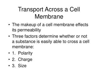
Transport: • Across tissue barriers • Gut Wall (absorption of nutrients) • Lung Alveoli (exchange of gases) • Capillary Beds (blood-tissue exchange of nutrients, gases, and waste products) • Nephrons in kidneys (urine formation) • Transcellular versus Paracellular • Across cell membranes • Regulation of cell size • Regulation of cell electrical activity • Passive vs. Active Transport
Example of transport across a tissue: Epithelium of small intestine Ganong, Fig. 25-7 Find: trancellular transport, paracellular transport
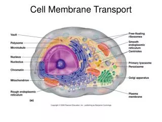
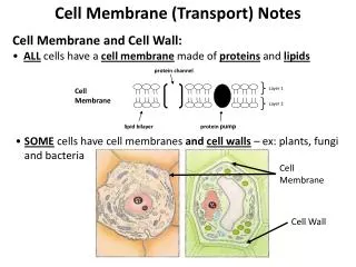
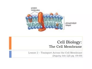
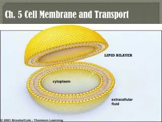
Cell Membranes: Fluid Mosaic Model Phospholipid bilayer, and integral membrane proteins Fig. 1-5 Ganong
Cell membranes: Selective Barriers • Lipophilic compounds can pass through. • Hydrophilic compounds do not pass through. • Specialized transporters and channels allow transport of hydrophilic compounds. • Channels - selective pores for ions, water, and very small molecules. Constructed from proteins. • Transporters - membrane proteins that move particular ions or small molecules across membranes.
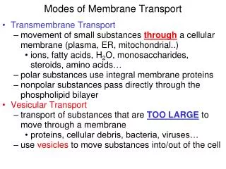
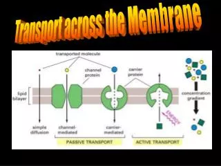
Transport across membranes: • Passive Transport • Diffusion, Osmosis • Solutes or water cross a membrane by following a concentration gradient. • Facilitated Diffusion • Carrier Molecule binds a solute and brings it across the membrane with no energy expended. • Active Transport • Requires energy expenditure, and can go against a gradient.
Diffusion: Fick’s First Law Js = -PS(Co - Ci) “Outside” “Inside” Js = solute flux; P = permeability coefficient; S = surface area for diffusion; (C0 - Ci) = concentration gradient of the solute
What determines permeability (P)? P = kT/6πrηx k = Boltzmann’s constant T = absolute temperature r = radius of the solute η = viscosity of the medium x = thickness of membrane
Osmosis: Transport of Water Water also moves down its concentration gradient. A B A B Osmotic pressure = amount of pressure to keep water from entering side B in this diagram.
Osmosis • Described by van’t Hoff Equation: • π = RT(ϕic) • π = osmotic pressure • R = ideal gas constant • T = absolute temperature • ϕ = osmotic coefficient • i = number of ions formed by dissociation of a solute molecule • c = molar concentration of a solute • (ϕic) = osmolarity of the solution
Molarity and Osmolarity • Molarity = number of molecules in solution (moles/L). • Osmolarity = number of particles in solution (osmoles/L). • So, for molecules that don’t dissociate in solution, molarity = osmolarity • For molecules that do dissociate in solution, osmolarity will be higher than molarity.
Osmolarity vs. Molarity • Examples: • Glucose - doesn’t dissociate in solution. If one mole is dissolved in one liter of water, you get 1 mol/L or 1 M. Osmolarity = 1 osmoles/L. • NaCl - dissociates into Na and Cl ions in solution, so, a 1 M solution has an osmolarity of 2 osmoles/L. • CaCl2 - dissociates into Ca and 2 Cl ions in solution, so a 1 M solution has an osmolarity of 3 osmoles/L.
How do permeating solutes affect osmotic flow? • Flow water (V) = “Permeability” of Water (L) x Pressure Gradient • L = hydraulic conductivity • The pressure gradient is directly proportional to π. (V = Lπ) when solutes are impermeable. • Correction with permeant solute:V = σLΔπ • σ = reflection coefficient, a value from 0 to 1 • 0 = freely permeable; 1 = impermeable
Modes of transport for ions and small molecules across biological membranes • Channels • Transporters • ATPases • Other - endocytosis (not really across membrane, but into vesicle inside cell)
Ligand-gated ion channel Voltage-gated ion channel Electrical recording of gating
Patch Clamping is an experimental technique to measure gating of ion channels. Fig. 1-28
Active Transport Example: Sodium-Potassium ATPase (Na/K Pump) Costs 1 ATP to transport 3 Na and 2 K Fig. 1-32
Transporters (Facilitated Diffusion) Selective for particular solutes Can be Reversible
Example of transport across a tissue: Epithelium of small intestine Ganong, Fig. 25-7 Find: trancellular transport, paracellular transport, passive diffusion, facilitated diffusion, active transport
Gap Junctions (Connexons) are specialized channels that allow direct movement of ions and water between cells Fig. 1-13
Cells also can pick up and release water and solutes using vesicles. Exocytosis Endocytosis Fig. 1-25
How are signals spread in neurons and muscle cells?
The composition of various transporters and channels on a cell determines membrane potential (Vm). Fig. 1-33
Typical Concentrations of ions inside and outside human nerve cells:
Ionic equilibria can produce electrochemical potential differences. A B 100 mM K+ 10 mM K+ Δμ(K+) = RTln([K+]A/[K+]B) + zF(EA - EB) = “diffusive” + “electrical” influences
Electrochemical equilibrium can be described by the Nernst Equation: At equilibrium, Δμ(K+) = 0, so at equilibrium: RTln([K+]A/[K+]B) = zF(EA - EB), or EA - EB = (RT/zF)ln([K+]A/[K+]B) solving for RT/F and converting ln to log, EA - EB = (-60mV/+1) x log([K+]A/[K+]B)
A B 100 mM K+ 10 mM K+ So for the above example, EA - EB = (-60mV/+1) x log([0.1 M]/[0.01 M]) EA - EB = -60 mV x log 10 EA - EB = -60 mV
What does this mean? To hold a 10-fold concentration difference of K+ across a membrane, an electrical potential difference of about 60 mV is required. Why is this important? These potential differences are an energy source transmission of signals in neurons and muscle cells.
Gibbs-Donnan Equilibrium • The cytoplasm contains many large proteins, which typically have a negative charge. This influences how cation-anion pairs distribute across the membrane, creating an uneven distribution. • These nondiffusible ions influence how diffusible ions distribute across a membrane, and is known as the Gibbs-Donnan Effect.
The composition of various transporters and channels on a cell determines membrane potential (Vm). Fig. 1-33
Resting Membrane Potential: Depends mainly on electrical potential differences generated by Na, K, and Cl across cell membrane. ENa = (-60mV/+1) x log([14 mM]/[142 mM] = +60 mV EK = (-60mV/+1) x log([140 mM]/[4 mM] = -93 mV ECl = (-60mV/-1) x log([9 mM]/[125 mM] = -68 mV The resting potential is estimated by a weighted average of the above potentials. Weight is based on a parameter called conductance (inverse of electrical resistance)
Actual resting potentials (Em) are about -70 mV. ENa = +60 mV, which is a 130 mV difference from Em This difference represents a potential driving force. Nerve and muscle cells use this to propagate signals.
Voltage Gated Ion Channels - open or closed depending on membrane potential Fig. 1-30
At a “threshold” potential, voltage-gated sodium channels open, causing rapid influx of sodium an driving membrane potential up (start of action potential)
Figure 15-23 Rhythmic waves of smooth muscle contraction in the gut are the result of waves of action potentials moving along via gap junctions.
Regulation of vascular smooth muscle tone: Role of calcium in E-C coupling Fig. 21-2, Berne & Levy
? Ca-Calmodulin MLC MLC Phosphatase MLC Kinase MLC-PP Activation of Actin-Myosin ATPase Actin-myosin cross bridge reaction Contraction
Types of smooth muscle contraction: • Tonic Contraction = Constrict or Relax • All Smooth Muscle Types • Phasic Contraction = Short contraction followed by period of relaxation • GI smooth muscle - mixing contractions - will cover in more detail in next lecture.