Download

1 / 36
400 likes | 599 Vues
Biology 624 - Developmental Genetics. Lecture #9 -Blood Vessel Formation. Blood vessel formation is important in: Embryogenesis Pregnancy Wound healing Disease. Tumor vascularization is a major step in tumor progression.

E N D
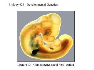
Biology 624 - Developmental Genetics Lecture #9 -Blood Vessel Formation
Blood vessel formation is important in: Embryogenesis Pregnancy Wound healing Disease
Diabetic retinopathy (excess blood vessels in the eye) is a major cause of blindness
VEGF Signaling Pathway FromShibuya, 2006
Cell Division + Migration + Survival + Cell Division - Migration + VEGF Signaling Pathway VEGF Flk-1 Flk-1 Flt-1 VEGF Angioblast Endothelial Cell
ES cell mutant vascular phenotype mimics mutant phenotype in vivo Wild-type Flk-1-/- Flt-1-/-
angioblasts Vascular development - differentiation of angioblasts Mesoderm Hemangioblast Hematopoietic cells
Embryonic vascular differentiation via the hemangioblast
angioblasts Vascular development - differentiation of angioblasts Mesoderm Hemangioblast Hematopoietic cells
Single cell fate mapping in fish gives Evidence for the hemangioblast FITC = descendants of single cell at 40% epiboly From Vogeli et al, 2006
angioblasts Embryonic vascular development Sprouting Angiogenesis Recruitment of mural cells and Remodeling Vasculogenesis proliferation migration VSMCs/pericytes differentiation assembly primitive vessel mature blood vessel branching vascular plexus
angioblasts Vasculogenesis VEGF bFGF differentiation assembly primitive vessel
Angiogenesis VEGF/neuropilin Slit/robo Netrin/unc Ephrin/eph Notch/delta proliferation migration sprouting branching vascular plexus primitive vessel
Retinal angiogenesis occurs outward from optic nerve postnatally From Gerhardt et al, 2003
Tip cell vs. stalk cell Tip cell: Does not proliferate Extends numerous filopodia Expresses markers- VEGFR2, PDGFB, Dll4, etc Stalk cell: Proliferates Has few filopodia Down-reglates tip cell markers From Gerhardt et al, 2003
From Gerhardt et al, 2003 Tip cell characteristics Express PDGFB No proliferation Actin-rich filopodia
VEGF pathway influences tip cells From Gerhardt et al, 2003
Cell Division + Migration + Survival + Cell Division - Migration + VEGF Signaling Pathway VEGF Flk-1 Flk-1 Flt-1 VEGF Angioblast Endothelial Cell Soluble flt-1 isoform selectively rescues migration/branching
Emerging flt-1 mutant sprouts differ from WT sprouts Local sprout guidance measures are rescued by sFlt-1 but not mFlt-1 Chappell et al. (2009) Dev Cell 17, 377
Local sprout guidance measures are perturbedIn retinas with reduced Flt-1 activity Flt-1 neutr ab flt-1flox/flox Adeno-Cre flt-1flox/flox flt-1flox/flox Chappell et al. (2009) Dev Cell 17, 377
Day 0 flt-1-/- ES cells Wild-type ES cells “Hanging drop” EBs Day 8 Mosaic blood vessels 1:1
Local sprout guidance measures are rescued by sFlt-1 but not mFlt-1 in lateral base areas Chappell et al. (2009) Dev Cell 17, 377
Local Sprout Guidance -- “pushes” the nascent sprout away from parent vessel -- utilizes soluble Flt-1 generated by parent vessel
Lumen formation in developingblood vessels HUVEC Zebrafish Kamei et al, 2006
Discontinuous lumens form and then connect in developingblood vessels Kamei et al, 2006
Intersomitic vessels appear composed of 4-7 cells and intercellular junctions Mosaic analysis Junction marker stain Blum et al, 2008
Putative structure of zebrafish intersomitic vessels Blum et al, 2008
The mouse DA forms via Cord Hollowing Strilic et al, 2009
Guidance pathways in vascular development From Klagsbrun and Eichmann, 2005
Remodeling Ang PDGF Recruitment of Smooth muscle /pericytes branching vascular plexus mature blood vessel
A-V formation in fish requires Vegfa Saggital sections of zebrafish over time show that Vegfa knockdown enhances CV formation at the expense of DA Herbert et al, 2009, Science Transverse sections show that Vegfa perturbations enhance CV formation at the expense of DA, while opposite for PI3K inh.
Blocking flow down-regulates arterial markers and up-regulates venous markers From le Noble et al, 2004