Download
1 / 17
200 likes | 706 Vues
C hronic lymphocytic leukemia. Magdalena Olszewska-Szopa. Definition. Chronic lymphocytic leukemia (CLL) belongs to B- cell low-grade non- Hodgkin lymphomas . CLL is the most common form of leukemia in adults. International Workshop on CLL (IWCLL) 2008 guidelines.

E N D
Chroniclymphocyticleukemia Magdalena Olszewska-Szopa
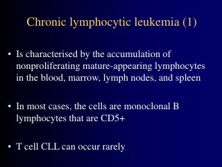
Definition Chroniclymphocytic leukemia (CLL) belongs to B-celllow-grade non-Hodgkinlymphomas. CLL is themost common form of leukemia in adults. International Workshop on CLL (IWCLL) 2008 guidelines
Epidemiology and riskfactors Riskfactors: • Advanced age • Positive family history
Clinicalpresentation • manypatientsareasymptomaticat the moment of the diagnosis, • fatigue, • autoimmunehemolytic anemia (AIHA), • infections, • splenomegaly, • hepatomegaly, • lymphadenopathy (painless), • extra-nodalinfiltrates – rarely.
Physicalexamination Lymphadenopathypresent in 87%: cervical/axillary/inguinal/generalised. Splenomegalypresent in 54% Hepatomegalypresent in 15% www.gp.online
Laboratory and imagingtests • Blood smear • Immunophenotyping – diagnosisconfirmation and riskfactorsidentificationeg ZAP70 and CD38 status • Interphase fluorescence in situ hybridization (FISH) - indetifiesgeneticriskfactorseg del17p • Serum marker beta-2 microglobulin • Concentration of immunoglobulins in the blood • Ultrasound of the abdomen and chestradiography • CT scans of the chest and abdomen (not mandatory) bloodtests -
Laboratory and imagingtests • Bone marrow tests usually are not needed. • Lymphnodebiopsyusuallyis not needed. Biopsiesareperformedif the peripheral blood smear does not yield diagnostic clues, to confirm the diagnosis, or to differentiate CLL from other diseases (e.g., Hodgkin's disease). Histopathologyorimmunophenotyping of lymphnodesareperformed in case of Richter syndrom suspicion.
Blood smear CLL cellsaretypically small/medium sizednormallookinglymphocytes Gumprechtshadows – smudgecells- typical for CLL ASH Image Bank - American Society of Hematology
Immunophenotyping – flowcytometry The test whichisusuallydone from blood- determineswhether the cellsareclonal and recognizestheirantigens (CD’s). Classicalfindings: CD19+, CD20weak +, CD23+, lightchainrestriction (kappa or lambda) AdverseFlow-cytometryfactors: High proportion of CLL cells containing ZAP-70 (20% or more) or CD38 (30% or more)
Rai staging and prognostic system: Leukemia and lymphomasociety (2017) Binetstaging system: Adapted from 2008 NCI guidelines
Assesingpatient fitness • Eastern Cooperative Oncology Group (ECOG) Performance Status • CumulativeIllness Rating Scale (CIRS)
Differentialdiagnosis • SLL – small lymphocyticlymphoma. The same appearance of neoplasticcells. The tissue infiltration issituatedin lymph nodes, spleen, or other organs but circulating B lymphocyte count <5 x 10⁹/L • NHL – other non hodgkinlymphomas. Histopathologicaltestsneeded. • Reactivelymphocytosis– the cellsare not clonal. • MBL – monoclonal B cel lymphocytosis. As many as 12% of healthy individuals >40 years of age may have low levels (<5 x 10⁹/L) of circulating monoclonal B cells with no evidence of organ infiltration. MBL progresses to CLL at a rate of 1%–2% of patients per year • Othertypes of chronić leukemias, eg: hairycell leukemia, prolymphocytic leukemia..
Prognosticfactors • Serum markers, such as beta2 –microglobulin • Geneticmarkers, including immunoglobulin heavy chainvariable region (IgHv), genemutational status (se: NOTCH1 genemutation, tp53 mutation) • Geneticabnormalitiesdetected by fluorescence in situ hybridization (FISH) ormetaphasecytogenetics (se: del 17p) • Protein markers, such as zeta-chain-associated protein kinase 70 ([ZAP]-70), cluster of differentiationCD38, orCD49d. • Lymphocytedoublingtimeshorterthan one year • Increased fraction of prolymphocytes (an early form of the lymphocyte) in the blood Leukemia and Lymphomasociety
Complications • Immunosuppression with subsequent infections (most common cause of death) • Autoimmune hemolytic anemia (AIHA) (of both the warm and cold agglutinin type) • ImmuneThrombocytopenic Purpura (ITP) • Richter's transformation or Richter's syndrome: transformation into a high-grade NHL (approx. 5% of CLL cases) • Secondarymalignancies
Treatment • Watch and waitstrategy- no benefits of early treatment for people with low-risk CLL have been shown. • When to Begin Treatment(whenatleast one of the aboveispresent): • Enlarging lymph nodes noted over a series of clinical exams • Enlarging liver and/or spleen noted over a series of clinical exams • Decreased red blood cell counts (hemoglobin less than 11g/dL) • Decreased platelet counts (platelets less than 100,000/uL) • Autoimmune anemia and/or thrombocytopenia poorly responsive to standard therapy • Lymphocyte doubling time of less than 6 months (only in patients with lymphocytes greater than 30g/L) • Presence of CLL B - symptoms
Therapy The therapymayconsist of classicaldrugs and immunotherapy: • Purineanalogues and alkylatingagents • Anti-CD20 antibodies • Glicocortycosteroidsare part of the regimensoraregiven in monotherapy to eliminateautoimmunologiccytopenias. Targetedtherapyis of increasingimportance: • Brutonkinaseinhibitors, • PI3K inhibitors, • Bcl-2 inhibitors
Therapy • Autologousstem cel translantation (ASCT) – due to the lack of efficacy and relatively high risk of latecomplications ASCT is NOT a part of therapy • Allogenicstem cel transplantation (Allo-SCT) – decreasing role in the era of newdrugs. Stillanoption for young high riskorrecurrentpatients. • Surgeryorradiotherapyhave no role in the therapy


![[PDF] DOWNLOAD Medifocus Guidebook on: Chronic Lymphocytic Leukemia](https://cdn7.slideserve.com/12581635/slide1-dt.jpg)