Thromboelastography
Thromboelastography. Dr. S. Parthasarathy MD., DA., DNB, MD ( Acu ), Dip. Diab . DCA, Dip. Software statistics PhD ( physio ) Mahatma Gandhi medical college and research institute , puducherry – India . What is it and when started .

Thromboelastography
E N D
Presentation Transcript
Thromboelastography Dr. S. Parthasarathy MD., DA., DNB, MD (Acu), Dip. Diab. DCA, Dip. Software statistics PhD (physio) Mahatma Gandhi medical college and research institute , puducherry – India
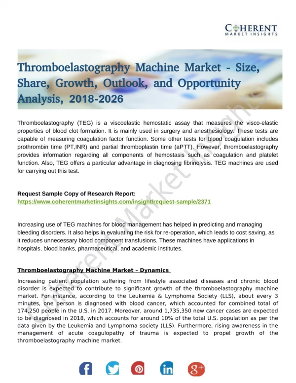
What is it and when started • Thromboelastographyis a point-of-care global hemostasis system used by anaesthetists largely to monitor perioperative changes in coagulation in surgical patients • It was first developed by the German Dr. HellmutHartert at University of Heidelberg School of Medicine. in 1948
Indications • Cardiac Surgery • Patient factors(aspirin,clopidogrel,heparin, clotting disorders) • Vascular Surgery. • Major obs. Haemorrhage • Regional on heparin • liver transplantation
What does it test ?? • The visco elastic changes that occur during the hemostaticprocess • TEG provides a real-time functional evaluation of the coagulation cascade, • Platelet function • Clot formation • clot strengthening and fibrin cross-linkage • clot lysis
classical thromboelastography • a small sample of blood is placed into a cuvette (cup) rotated gently to imitate sluggish venous flow and activate coagulation. • When a sensor shaft is inserted into the sample a clot forms between the cup and the sensor. • The speed and strength of clot formation is measured depends on the activity of the plasmatic coagulation system, platelet function, fibrinolysis and other factors
Values • the R value - time until the first evidence of a clot is detected • The K value - the end of R until the clot reaches 20mm and this represents the speed of clot formation • The angle is the tangent of the curve made as the K is reached and offers similar information to K • The MA is a reflection of clot strength. • Clot lysis time
R value • R: R time is the period of latency from the time that the bloodwas placed in the TEG analyzer until the initial fibrin formation.This represents the enzymatic portion of coagulation. (clotting factors ) 4 – 8 minutes • R = 11 – 14 min – FFP 8 ml/kg • R > 14 FFP 15 ml/Kg
Prolonged R • Shortened R • Hypercoagulable state and early DIC
K: K time is a measure of the speed to reach a certain levelof clot strength. This represents clot kinetics. • 1 – 4 minutes • alpha : measures the rapidity of fibrin built up and cross-linking(clot strengthening). This represents fibrinogen level. • 45 – 75 deg
R normal • K prolonged • Platelet defect
MA: MA or maximum amplitude, is a direct function of the maximumdynamic properties of the fibrin and platelet bonding via GPIIb/IIIaand represents the ultimate strength of the fibrin clot. • 55 – 70 mm • 80 % platelets • 20 % fibrinogen
Decrease and continuous decrease • Platelet deficiency • Primary fibrinolysis
LY30: LY30 measures the rate of amplitude reduction 30 minafter MA. This represents clot lysis. • Normal = 7.5 %
TO SUMMARIZE • Cuvette pin 0.3 ml blood • Visco elastic properties • Full coagulation profile • R, K ,MA , LY 30 • Disease s
TEG with heparinase • heparinase (an enzyme breaking heparin) • “heparinized” patients, prior to heparin reversal with protamine. • This test may prove particularly useful during long pump runs, deep hypothermia, use of ventricular assist devices or complicated major vascular cases • The test will also detect if more protamine is needed to fully reverse heparin.
To remove platelet effect • Add recently FDA approved monoclonal antibody which binds to the platelet GPIIb/IIIa receptors, to the TEG sample • eliminate platelet function from the thromboelastogram • The MA will become a function of fibrinogen activity
The Sonoclot • provides an alternative visco elastic measure of coagulation. • In contrast to the TEG, Sonoclot immerses a rapidly vibrating probe into a 0.4-mL sample of blood. • As clot formation occurs, impedance to probe movement through the blood increases and generates an electrical signal and characteristic clot “signature.” • The Sonoclot may be used to derive the ACT, as well as to provide information regarding clot strength and fibrinolysis.
Treat the patient than TEG • Thank you all