Download

1 / 125
1.71k likes | 3.46k Vues
Vocal cord palsy & evaluation of hoarseness. Dr. Vishal Sharma. Nerve supply of larynx. Motor supply of intrinsic muscles: Cricothyroid muscle: superior laryngeal nerve All other muscles: recurrent laryngeal nerve Sensory: Above vocal cord: superior laryngeal nerve

E N D
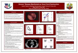
Vocal cord palsy & evaluation of hoarseness Dr. Vishal Sharma
Nerve supply of larynx Motor supply of intrinsic muscles: Cricothyroid muscle:superior laryngeal nerve All other muscles:recurrent laryngeal nerve Sensory: Above vocal cord:superior laryngeal nerve Below vocal cord:recurrent laryngeal nerve
Recurrent laryngeal nerve Right: • Arises from vagus at level of right subclavian artery & hooks around it Left: • Arises from vagus in mediastinum at level of arch of aorta & loops around it
Superior laryngeal nerve • Arises from inferior ganglion of vagus • Descends behind internal carotid artery at level of greater cornu of hyoid bone divides into external & internal branches • External motor branch:to cricothyroid muscle • Internal sensory branch:pierces thyrohyoid membrane to enter larynx
Classification A. Incomplete paralysis 1. Recurrent laryngeal nerve palsy a. Left (75% ), Right (15%), B/L (10%) b. Abductor, Adductor 2. Superior laryngeal nerve palsy B. Combined paralysis / complete paralysis
Causes of laryngeal paralysis Supra-nuclear Nuclear:nucleus ambiguus High vagal lesions:combined palsy Low vagal lesions: recurrent laryngeal nerve palsy Systemic causes Idiopathic
Causes of combined paralysis IntracranialNeck Tumors of posterior fossaPenetrating injury Basal meningitis (TB) Parapharyngeal tumors Skull baseMetastatic neck nodes Fractures Lymphoma Nasopharyngeal cancer Thyroid surgery Glomus tumour
Malignancy (25%):lung (>50%), thyroid, esophageal, nasopharyngeal, metastatic neck node Surgical trauma (20%):during surgeries of lung, heart, thyroid, esophagus, mediastinum Inflammatory (13%):tuberculosis, syphilis Idiopathic (13%):viral neuritis Non-surgical trauma (11%):accidental neck trauma, left atrial enlargement (Ortner), aortic aneurysm Neurological (7%):CVA, head injury, Parkinsonism, multiple sclerosis, alcoholic / diabetic neuropathy Others (11%):rheumatoid arthritis, haemolytic anemia
Neck Accidental trauma Thyroid disease Thyroid surgery Ca esophagus Lymphadenopathy Mediastinum Bronchogenic ca Ca esophagus Aortic aneurysm Lymphadenopathy Enlarged left atrium Intra-thoracic surgery Causes of left RLN palsy (75%)
Causes of right RLN palsy (15%) • Neck trauma • Thyroid disease • Thyroid surgery • Ca cervical esophagus • Cervical lymphadenopathy • Aneurysm of subclavian artery • Ca apex right lung • TB of cervical pleura
Causes of B/L RLN palsy (10%) • Thyroid surgery • Ca thyroid • Cancer cervical esophagus • Cervical lymphadenopathy
Congenital vocal cord paralysis Unilateral:birth trauma, congenital anomaly of great vessel or heart Bilateral: Hydrocephalus Meningocoele Arnold-Chiari malformation Cerebral agenesis Intra-cerebral hemorrhage Nucleus ambiguus agenesis
Thyroid surgery Joll’s sterno-thyro-laryngeal triangle for S.L.N.: Lateral = superior thyroid vessels & upper thyroid pole; superior = attachment of strap muscles to thyroid cartilage; medially = midline Beahr’s triangle for recurrent laryngeal nerve: Lateral = common carotid artery; superior = inferior thyroid artery; medial = tracheo-esophageal groove + recurrent laryngeal nerve
Why right RLN commonly damaged in thyroid surgery? • Right recurrent laryngeal nerve more superficial • Right nerves enters thyroid at 450 angle but left lies in tracheo-esophageal groove • Right nerve mostly passes superior to or b/w branches of inferior thyroid artery; left nerve mostly passes deep to inferior thyroid artery
Semon’s Law Rosenbach (1880) & Semon (1881) “In all progressive organic lesions, abductor fibres of recurrent laryngeal nerve, which are phylogenetically newer, are more susceptible and thus first to be paralyzed compared to adductor fibres.”
1st stage:only abductor fibres damaged; vocal folds approximate in midline; adduction still possible (paramedian position) 2nd stage:contracture of adductors; vocal folds immobilized in median position 3rd stage:adductors become paralyzed; vocal fold assumes cadaveric position
Why abductors affected first ? • Nerve fibres supplying abductors are in periphery of recurrent laryngeal nerve • Muscle bulk for the abductors is less, more susceptible • Phylogenetically, larynx’s main function is protection, so adductor functions are maintained
Wagner & Grossman Theory In isolated paralysis of recurrent laryngeal nerve, cricothyroid muscle (which receives innervation from superior laryngeal nerve) keeps vocal cord in paramedian position due to adductor function In superior laryngeal nerve palsy, cord lies in intermediate (cadaveric) position
Modern theory Final position of paralyses vocal cord is not static & is decided by: • Degree of paralyzed muscle atrophy& fibrosis • Degree of re-innervation following injury • Extent of synkinesis (mass movement) of all intrinsic muscles • Fibrosis & ankylosis of crico-arytenoid joint
Intermediate position of vocal cords in RLN palsy? Retrograde atrophy of vagus nerve occurs up to nucleus ambiguus Stretching of RLN by enlarged intra-thoracic lesions pulls vagus nerve down from skull base, injuring superior laryngeal nerve
Lesion above pharyngeal branch • Inability to elevate soft palate, nasal intonation, nasal regurgitation & nasal emissions • Gag reflex reduced or absent due to palsy of internal branch of superior laryngeal nerve • Hoarseness due to palsy of intrinsic muscles of larynx
Voice assessment 1. Magnetic tape recording:for self assessment 2. Performance assessment by examiner: maximum phonation time & range of speech frequencies 3. Phonetogram:plot of pitch vs. intensity of voice 4. Aerodynamic analysis: phonatory airflow rate, subglottic pressure & laryngeal resistance
5. Fourier’s Spectral analysis (Spectrogram) • Fundamental frequency:lowest speech frequency • Shimmer:average cycle to cycle difference in amplitude of sound • Jitter:average cycle to cycle difference in duration of glottal cycle In hoarseness there is increased shimmers & jitters
Analysis of cord movement 1. Rigid 700 video-telescopy ↓LA 2. Fibreoptic video-laryngoscopy 3. Stroboscopy: Intermittent flash light focussed on vocal cords during phonation. Frequency of light made 2 msec slower to cord frequency. Produces slow motion movement of vocal cords for better analysis of cord movement
4. Electro-glottography: 2 electrodes placed on both sides of thyroid cartilage & current passed b/w them. Recorded waveform shows impedance across larynx & is highest during contact b/w vocal cords. Records closing phase of glottal cycle. 5. Photo-glottography:fibreoptic light source passes light via glottis & is received by photo-sensor on neck skin. Light received glottic chink. Records opening phase of glottal cycle.
Radiological • Submento-vertical skull base view • X-ray neck AP & lateral view • Chest X-ray PA view • Barium swallow AP & lateral oblique view • High resolution CT scan with contrast from skull base to mid thorax:gold standard • M.R.I.:ideal for skull base lesions • Thyroid scan
Endoscopy 1. Rigid 700 Telescopy ↓ LA 2. Fibreoptic Laryngoscopy ↓ LA 3. Pan-endoscopy ↓ GA (for metastatic node): a. Nasopharyngoscopy b. Micro-laryngoscopy: probe test on arytenoids c. Bronchoscopy & bronchial washings d. Hypopharyngoscopy e. Oesophagoscopy