Download
1 / 1
10 likes | 110 Vues
Crackle pitch rises progressively during inspiration in pneumonia, CHF and IPF patients Raymond Murphy and Andrey Vyshedskiy, Brigham and Women’s / Faulkner Hospitals, Boston MA. Highest peak. Introduction. A1. A 2.

E N D
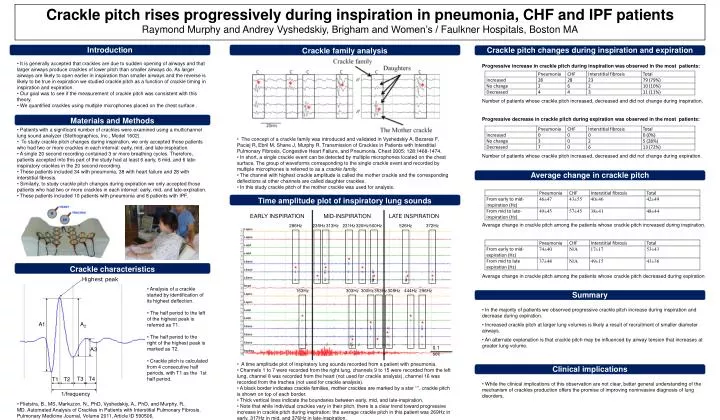
Crackle pitch rises progressively during inspiration in pneumonia, CHF and IPF patients Raymond Murphy and Andrey Vyshedskiy, Brigham and Women’s / Faulkner Hospitals, Boston MA Highest peak Introduction A1 A2 • It is generally accepted that crackles are due to sudden opening of airways and that larger airways produce crackles of lower pitch than smaller airways do. As larger airways are likely to open earlier in inspiration than smaller airways and the reverse is likely to be true in expiration we studied crackle pitch as a function of crackle timing in inspiration and expiration. • Our goal was to see if the measurement of crackle pitch was consistent with this theory. • We quantified crackles using multiple microphones placed on the chest surface . Progressive increase in crackle pitch during inspiration was observed in the most patients: A3 Number of patients whose crackle pitch increased, decreased and did not change during inspiration. T3 T4 T1 T2 Progressive decrease in crackle pitch during expiration was observed in the most patients: 1/frequency • Patients with a significant number of crackles were examined using a multichannel lung sound analyzer (Stethographics, Inc., Model 1602). • To study crackle pitch changes during inspiration, we only accepted those patients who had two or more crackles in each interval: early, mid, and late-inspiration. • A single 20 second recording contained 3 or more breathing cycles. Therefore, patients accepted into this part of the study had at least 6 early, 6 mid, and 6 late-inspiratory crackles in the 20 second recording. • These patients included 34 with pneumonia, 38 with heart failure and 28 with interstitial fibrosis. • Similarly, to study crackle pitch changes during expiration we only accepted those patients who had two or more crackles in each interval: early, mid, and late-expiration. • These patients included 10 patients with pneumonia and 8 patients with IPF. 0.1 sec Crackle family analysis Time amplitude plot of inspiratory lung sounds Crackle characteristics Materials and Methods • The concept of a crackle family was introduced and validated in Vyshedskiy A, Bezares F, Paciej R, Ebril M, Shane J, Murphy R. Transmission of Crackles in Patients with Interstitial Pulmonary Fibrosis, Congestive Heart Failure, and Pneumonia. Chest 2005; 128:1468-1474. • In short, a single crackle event can be detected by multiple microphones located on the chest surface. The group of waveforms corresponding to the single crackle event and recorded by multiple microphones is referred to as a crackle family. • The channel with highest crackle amplitude is called the mother crackle and the corresponding deflections at other channels are called daughter crackles. • In this study crackle pitch of the mother crackle was used for analysis. Number of patients whose crackle pitch increased, decreased and did not change during expiration. Clinical implications Crackle pitch changes during inspiration and expiration Average change in crackle pitch Summary EARLY INSPIRATION MID-INSPIRATION LATE INSPIRATION Average change in crackle pitch among the patients whose crackle pitch increased during inspiration. 286Hz 235Hz 313Hz 231Hz 320Hz 500Hz 526Hz 372Hz * * * * * * * * Average change in crackle pitch among the patients whose crackle pitch decreased during expiration • Analysis of a crackle started by identification of its highest deflection. • The half period to the left of the highest peak is referred as T1. • The half period to the right of the highest peak is marked as T2. • Crackle pitch is calculated from 4 consecutive half periods, with T1 as the 1st half period. 163Hz 303Hz 300Hz 353Hz 308Hz 444Hz 296Hz • In the majority of patients we observed progressive crackle pitch increase during inspiration and decrease during expiration. • Increased crackle pitch at larger lung volumes is likely a result of recruitment of smaller diameter airways. • An alternate explanation is that crackle pitch may be influenced by airway tension that increases at greater lung volume. * * * * * * * • A time amplitude plot of inspiratory lung sounds recorded from a patient with pneumonia. • Channels 1 to 7 were recorded from the right lung, channels 9 to 15 were recorded from the left lung, channel 8 was recorded from the heart (not used for crackle analysis), channel 16 was recorded from the trachea (not used for crackle analysis). • A black border indicates crackle families, mother crackles are marked by a star ‘*’, crackle pitch is shown on top of each border. • Thick vertical lines indicate the boundaries between early, mid, and late-inspiration. • Note that while individual crackles vary in their pitch, there is a clear trend toward progressive increase in crackle pitch during inspiration: the average crackle pitch in this patient was 269Hz in early, 317Hz in mid, and 376Hz in late-inspiration. • While the clinical implications of this observation are not clear, better general understanding of the mechanism of crackles production offers the promise of improving noninvasive diagnosis of lung disorders. • Flietstra, B., MS, Markuzon, N., PhD, Vyshedskiy, A., PhD, and Murphy, R., MD. Automated Analysis of Crackles in Patients with Interstitial Pulmonary Fibrosis. Pulmonary Medicine Journal, Volume 2011, Article ID 590506,