Download
1 / 47
490 likes | 1.01k Vues
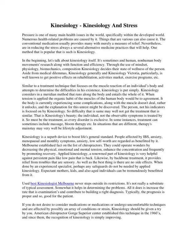
Objectives. Introduction to applied kinesiologySimple tests to implement in your practiceDiagnosis of spheno-basilar fixationRespiratory cranial faults recognitionSutural cranial fault detectionCautions. 2. 3. Introduction to kinesiology. History

E N D
1. THE USE OF KINESIOLOGY IN DIAGNOSING CRANIAL FAULTS
Richard Cook DC,FCC,CCEP.
Sat. 17th April 2010
2. Objectives Introduction to applied kinesiology
Simple tests to implement in your practice
Diagnosis of spheno-basilar fixation
Respiratory cranial faults recognition
Sutural cranial fault detection
Cautions 2
3. 3
4. Introduction to kinesiology History � kinesiology was discovered in 1964 by US chiropractor Dr. George Goodheart.
Why would fit people have muscle weaknesses?
Muscles can be tested for strength or weakness
An organ-muscle relationship
Muscle testing procedures
�The truth is out there!�
The body never lies 4
5. Grading muscle strength 5
6. Kinesiology �5 minutes to learn yet a lifetime to master�
Uses standard muscle testing procedures to provide useful information on the status of the body
Can be used to ask the body questions in a binary fashion receiving a �Yes� or �Not yes� response 6
7. Normal physiology A normal muscle is facilitated - that is on (strong)
All muscles in the body have another that does the opposite action � agonist/antagonist
When a muscle fails to work normally it is off (weak)
This is not a weakness that is associated with lack of exercise
We are testing functional neurology 7
8. Muscle testing Changes are instantaneous, repeatable and consistent.
They are mediated via the nervous system
In medicine muscle spasm is the priority
With kinesiology we look at the weakness as the primary problem 8
9. Muscle weakness Muscles can become weak for a multitude of reasons;
Neurolymphatic
Neurovascular
Nerve
Acupuncture � meridian/organ
Nutrition
Together these factors are known as the 5 factors of the intervertebral foramina 9
10. Advantages of kinesiology It shows you the priority
It can show you where to go next
It tells you which direction or respiratory phase in which to adjust
It will inform you that the job has been done 10
11. 11
12. Two ways to use ak Use a strong, normal indicator muscle
Discover by therapy localisation what weakens it
Use a related muscle that is weak in the clear
Find out what will restore it back to strength 12
13. 42 muscles There are 42 muscles which are on both sides of the body which are routinely tested
These relate to the 12 acupucture meridians and their organs kinesiology 13
14. 4 Bilateral arm muscle tests Pectoral major clavicular
Anterior deltoid
Latissimus dorsi
Supraspinatus 14
15. Why use both sides at once? The 4 muscles relate to:
Emotions
Structure
Body chemistry
Cranials
Also using both sides involves both brain hemispheres and that reduces the chances of the patient cheating!
15
16. The supraspinatus Supraspinatus is related to the brain
Generally muscles are tested individually
Sometimes we test muscles bilaterally
This takes out some of the ability to cheat! 16
17. The supraspinatus test Like every muscle test check first each independently
Idea is for the patient to bring origin and insertion of the muscle towards one another
Push gently asking the patient to resist the pressure
Continue the pressure for 2 seconds
Feel for a weakening or the ability to hold 17
18. Factors that may influence the test Watch the patient does not initiate the test by pushing first
Look for breath holding
Subtle changes in position of the limb being tested
The patient gritting the teeth
Pain on testing
18
19. All muscles test strong! Every muscle should be able to be switched off
This is done by pressing the appropriate sedation point on the related meridian
There may be hypertonicity in the first muscle you test if that is the case use another
When a weakening is difficult to locate it could be:-
An atlas fixation
TMJ problem
Cranial SBS jamming
Pelvic category problem 19
20. All muscles test weak! Dehydration is the most likely reason
The patient could fail to understand the instructions
There may be a debilitating disease present
Patient may have eyes closed 20
21. Why are cranials so important? 90% of the nervous system is above the atlas!!
The cranium is the box the brain comes in
Cranial faults can impinge directly on the brain and thereby influence remote function
In any chronic condition check the cranium
In virtually any problem look to the cranium first 21
22. 22
23. Background to cranials History is not relevant at this juncture.
There are many techniques and schools of thought
Soft v Hard?
I believe gentle is best as we are essentially
moving fluids.
Consider the surface anatomy of the head. 23
24. First contact!
The first time you touch the patient�s head is so important.
Move in gently and respectfully.
Wait to feel the sensations under your fingertips �
Rhythm frequency quality symmetry amplitude. 24
25. Every skull is unique
Each skull is a new learning experience
Lack of motion leads to reduced function
Symmetrical motion is better than asymmetrical
Balance is better than imbalance 25
26. Less is more?
Lighten up!
Enhance your palpatory skills.
Keep it soft and gentle and let the body do the work
26
27. Flexion - extension 27
28. Sheno-basilar fixation The spheno-basilar symphysis can become jammed thus not moving in the usual respiratory pattern
SBS compression is fairly common and can be the result of :-
Trauma
Fatigue
Stress
Birth trauma
Toxicity 28
29. Cranial Faults Conditions requiring cranial investigation Part 1
Cranial nerve involvement
Squint
Eye tracking problems
Bell�s palsy
Pituitary functional disturbance � hormonal problems
Trauma - head injury, whiplash
Malocclusion � look in the mouth! 29
30. Cranial Faults Conditions requiring cranial investigation Part 2
Birth trauma � restless infant, breathing problems, behavioural problems, vomiting, hypertonicity, tremor, and specific learning difficulties
Dural torque
Allergy
Infection
Toxicity
Emotional stress
Remote structural problems � pelvis, neck, spine, feet
30
31. Respiratory cranial flow chart 31
32. Sutural cranial flow chart 32
33. Cautions There may be another priority
Short cuts don�t always work
Never perform cranial work on a recent stroke victim
Never work on somebody with a suspected
skull fracture 33
34. Flow charts Respiratory cranial faults are altered by a phase of respiration
Sutural cranial faults are detected by TL
The sphenoid articulates with 12 other cranial bones and as such is viewed as the king pin of the geared system and should be checked in all patients 34
35. Palpating the cranial rhythm Skull bones are the handles that move and relieve tension in the dural membranes
Wait!
Feel beyond the obvious for-
Speed
Quality
Amplitude
Rhythm
Symmetry 35
36. Passion We all must get passionate about the cranial system
We need to get this information out there
We must be the therapists of choice, nobody else has the palpatory skills 36
37. 37
38. Thank you for your kind attention
�Nunc est bibendum�
Which roughly translated means �I�ve finished let�s go to the pub!� 38
39. 39
40. Let�s get physical! AK testing procedures �
Know your anatomy
Think what action the muscle performs
Origin and insertion move towards one another
Take care in your and the patient�s positioning 40
41. AK test Inform the patient what you are going to do
Wait for the patient to be ready
Push gently but firmly in the opposite direction to the muscle action
Push with a gradually increasing pressure for about 2 seconds
Feel for any weakening, give, juddering or shakiness
Repeat on the opposite side for comparison
41
42. What to avoid Pushing too hard
Pushing too early to beat the patient
Make sure the patient is competent and understands the test procedure
Don�t allow the patient to �
change the position of their limb being tested
Grit their teeth
Hold the breath
Cheat!
42
43. A tip
Always compare the other side where practicable.
Try to get an impression of each person�s
overall strength, by checking a few different muscles. 43
44. Rapid screen testing 5 limb individual tests a weakness indicates:
Neck flexors = C2-C7 subluxation
General leg = Category 2
Anterior deltoid = Shoulder problem
Psoas independently � both weak = atlas/occiput fixation 44
45. 4 Bilateral arm muscles Anterior deltoids related to Structural problems
Pect. major clav. � Emotional issues
Lat. Dorsi � Blood chemistry
Supraspinatus � Cranial fault 45
46. Supraspinatus Inability to hold bilaterally tested supraspinatus muscles that are independently strong indicates a
Spheno-basilar fixation.
Check whether insp or exp strengthens
To clear hold cranium in a vault hold and follow the patient�s deep respiration through 4-6 cycles 46
47. The beauty of AK When you have done something you can check to see
is there a change?
If the muscles now work fine the job is complete
If not you may have to repeat the procedure
Remember we are looking at functional neurology and not how strong the patient happens to be. 47
48. The body never lies! This is what Dr. George Goodheart quoted on many occasions and providing you are careful this is true.
However, the body will try and fool you when it can,
but treat that as a learning experience.
Any questions? 48