Download

1 / 51
570 likes | 790 Vues
Anatomy of the Hand. Goal. To discuss the basic anatomy of the hand. Terminology. Hand Dorsal surface Volar or palmar surface Radial and ulnar borders Palm Thenar area Mid-palm area Hypothenar area. Palmar view of the hand. Essentials of Hand Surgery 2002. Terminology. Digits

E N D
Goal • To discuss the basic anatomy of the hand
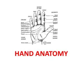
Terminology • Hand • Dorsal surface • Volar or palmar surface • Radial and ulnar borders • Palm • Thenar area • Mid-palm area • Hypothenar area Palmar view of the hand Essentials of Hand Surgery 2002
Terminology • Digits • Thumb, index, middle or long, ring, & small digits • Each finger has three joints • Metacarpophalangeal MCP), proximal interphalangeal (PIP), & distal interphalangeal (DIP) joints • Thumb • Two phalanges & two joints
Surface Anatomy Essentials of Hand Surgery 2002
Hand Motion • Standardized description • Forearm • Wrist • Digits • Center of hand • Long finger ray Essentials of Hand Surgery 2002
Thumb Motion • Standardized description • Palmar abduction • Planar abduction • Opposition • Abduction • Flexion • Pronation Essentials of Hand Surgery 2002
Palmar Fascia • Provides a stable platform for the palmar skin & protects the underlying structures • Insertion site for the palmaris longus tendon Essentials of Hand Surgery 2002
Palmar Spaces • Septum extends from the palmar aponeurosis to the third metacarpal • Thenar space located to the radial side of the septum • Midpalmar space situated on the ulnar side • Spaces can be infected primarily or after flexor tenosynovitis of the digits
Spaces Transverse section demonstrating septum from palmar fascia to third metacarpal, which divides hand into thenar and midpalmar space Essentials of Hand Surgery 2002
Blood Supply • Radial & ulnar arteries supply blood to the hand through a series of two arches Essentials of Hand Surgery 2002
Blood Supply-Radial Artery • Radial artery splits into two branches, with the larger dorsal branch coursing under the first dorsal compartment, through the anatomic snuffbox, between the index & thumb metacarpals, and into the recesses of the palm to form the greater part of the deep palmar arch
Blood Supply-Radial Artery • A smaller palmar branch travels over the flexor carpi radialis tendon, beneath or through the thenar muscles, and forms the radial component of the superficial palmar arch
Blood Supply- Ulnar Artery • Ulnar artery located medial to the ulnar nerve at the wrist & adjacent to flexor carpi ulnaris tendon • Ulnar nerve & artery course • into Guyon’s canal • Ulnar artery and nerve are covered by the volar carpal ligament in Guyon’s canal Essentials of Hand Surgery 2002
Blood Supply- Ulnar Artery • Ulnar artery splits into two branches with the larger branch forming the main constituent of the superficial palmar arch • Smaller branch passes deep to connect with the radial artery and to form the deep palmar arch
Blood Supply • A line drawn across the palm parallel to the fully abducted thumb (a.k.a. Kaplan’s cardinal line) approximates the location of the superficial palmar arch • This arch is located just past the distal edge of the TCL • The deep palmar arch is located 1 cm proximal to the superficial palmar arch & is beneath the flexor tendons
Blood Supply • Although considerable variability exists, the superficial palmar arch typically provides palmar blood vessels to the index (ulnar side), long, ring, and small fingers while the deep palmar arch supplies blood vessels to the thumb and index digit (radial side)
Blood Supply- Fingers • Common digital arteries originate from the superficial palmar arch & travel within the index-long, long-ring, & ring-small interspaces • Common digital arteries divide into proper digital arteries that continue along the side of the digit
Blood Supply- Thumb • The vascular supply to the thumb is from digitalbranches that originate as a common trunk (a.k.a. princeps pollicis artery) from the deep palmar arch Essentials of Hand Surgery 2002
Nerves • Radial, median, & ulnar nerves supply the sensibility and motor to the hand and forearm Courtesy of Scott H. Kozin, MD
Nerves- Median • Median travels down forearm between the flexor digitorum superficialis & profundus muscles to enter the carpal tunnel • Along its course, the anterior interosseous nerve branches to provide innervation to the flexor pollicis longus, flexor digitorum profundus to the index, & the pronator quadratus muscles
Nerves- Median • Recurrent motor branch originates from the central or radial portion of the median nerve during its passage through the carpal tunnel • The recurrent branch usually passes distal to the transverse carpal ligament (TCL) to innervate the thenar muscles. Uncommonly, the nerve can pass through the TCL (5-7% individuals)
Nerves- Ulnar • Ulnar nerve resides medial to the ulnar artery within Guyon’s canal • Provides a motor branch to the hypothenar muscles • Deep motor branch passes around the hook of the hamate to innervate the interossei, ulnar lumbricals, flexor pollicis brevis (deep head), and the adductor pollicis muscles
Muscle/ Tendon • The muscles and tendons about the hand originate in the • Forearm (extrinsic) or • Within the hand itself (intrinsic)
Muscle/ Tendon- Dorsal • Extrinsic extensor muscles generate tendons that align within compartments along the dorsum of the wrist • The extensor retinaculum is a two-centimeter band that extends form the radial to the ulnar side of the wrist. • Retinaculum is a strong pulley that holds the tendons close to the underlying bone
Muscle/ Tendon- Dorsal • Compartments • 1st- APL & EPB • 2nd- ECRL & ECRB • 3rd- EPL • 4th- EIP & EDC • 5th- EDQ • 6th- ECU Essentials of Hand Surgery 2002
Muscle/ Tendon- Palmar • Carpal tunnel contains the flexor apparatus and the median nerve at the wrist Essentials of Hand Surgery 2002
Muscle/ Tendon- Palmar • Carpal tunnel- contents • Median nerve • FDS (4), FDP (4), FPL • Long & ring FDS tendons lie more palmar than the index and small FDS tendons
Muscle/ Tendon- Palmar • Primary wrist flexors do not reside within the carpal tunnel • FCR travels in an adjacent separate fibrosseous tunnel • FCU inserts into the pisiform and distally into the fascia that is palmar to the carpal tunnel
Muscle/ Tendon- Hand • Thenar muscles • Opponens pollicis • Flexor pollicis brevis (FPB) • Abductor pollicis • All innervated by recurrent branch of the median nerve • FPB muscle receives dual innervation: • Recurrent branch of the median nerve • Deep motor branch of the ulnar nerve
Muscle/ Tendon- Hand • Hypothenar muscles • Opponens digiti quinti • Flexor digiti quinti • Abductor digiti quinti • All innervated by the ulnar nerve
Muscle/ Tendon- Hand • Muscles within the hand • Interossei, lumbricals, and adductor pollicis
Muscle/ Tendon- Hand • Four bipennate dorsal interossei that originate from adjacent sides of the metacarpals Essentials of Hand Surgery 2002
Muscle/ Tendon- Hand • Provide abduction of the finger along with MCP joint flexion & interphalangeal joint extension Essentials of Hand Surgery 2002
Muscle/ Tendon- Hand • Three smaller unipennate palmar interossei that originate from a single metacarpal Essentials of Hand Surgery 2002
Muscle/ Tendon- Hand • Provide adduction toward the long finger along with MCP joint flexion & interphalangeal joint extension Essentials of Hand Surgery 2002
Muscle/ Tendon- Hand • Four lumbrical muscles originate within the hand from the radial side of the flexor digitorum profundus tendons • Course beneath the intermetacarpal ligament & contribute to the lateral bands
Muscle/ Tendon- Hand • Adductor pollicis muscle similar to a palmar interosseus in terms of function • Originates from the third metacarpal & inserts into the base of the proximal phalanx and extensor apparatus • Contraction produces an intrinsic response with MCP joint flexion, interphalangeal joint extension, & thumb adduction
Flexor Tendons • Enter a fibro-osseous tunnel (flexor sheath) at the level of the MCP joint • Sheath thickened to produce strong annular pulleys that position the tendons close to the underlying bone • A2 and A4 pulleys are essential to prevent bowstringing
Flexor Tendons • Cruciate pulleys are interdigitated between the annular pulleys • Less critical for function • More flexible than the annular pulleys • During digital flexion & extension, cruciate pulleys to compress and stretch to allow allow full motion.
Flexor Tendons Annular and Cruciate Pulleys Essentials of Hand Surgery 2002
Extensor Apparatus • Extensor digitorum comminus tendons pass over MCP joint & are held in position by the sagittal hood Regional Review Course 1998
Extensor Apparatus • Distal to the MCP joint, the extensor digitorum comminus divides into three parts. The central portion continues distally and attaches to the base of the middle phalanx (a.k.a. central slip) Regional Review Course 1998
Extensor Apparatus • Lateral bands formed primarily by the tendons of the interossei and lumbricals • Lateral bands terminate at the base of the distal phalanx & are responsible for PIP & DIP joint extension Regional Review Course 1998
Osseous/ Ligaments • Carpus or wrist consists of eight bones organized into two rows Essentials of Hand Surgery 2002
Osseous/ Ligaments • Proximal row • Scaphoid, lunate, and triquetrum • Distal row • Trapezium, trapezoid, capitate, and hamate • Pisiform • Sesamoid bone within the FCU & does not participate in carpal motion
Osseous/ Ligaments • Bones within each row held together via interosseus ligaments • Integrity of these ligaments is critical to normal wrist motion (a.k.a. carpal kinematics)
Osseous/ Ligaments • Wrist also possesses ligaments that connect radius to carpus • Extrinsic ligaments • Palmar extrinsic ligaments more important to wrist stability & motion than dorsal ligaments Essentials of Hand Surgery 2002
Osseous/ Ligaments • Triangular fibrocartilage complex (TFCC) originates from the radius & attaches to the base of the ulnar styloid Courtesy of Scott H. Kozin, MD
Osseous/ Ligaments • Dorsal and volar radioulnar ligaments are thickenings of TFCC & provide stability to the distal radioulnar joint Courtesy of Scott H. Kozin, MD