Download

1 / 48
500 likes | 567 Vues
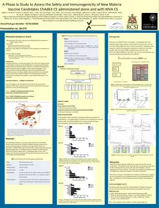
Learn about the definition, morphology, and life cycle of Plasmodia parasites causing malaria in humans. Understand the sexual and asexual cycles, identification features, and transmission methods. Study the infectious stages and symptoms caused by different Plasmodium species.

E N D
Plasmodium & Babesiosis Dr. Sameer Naji, MB, BCh, PhD (UK) Assistant Professor Dept. of Basic Medical Sciences Faculty of Medicine The Hashemite University
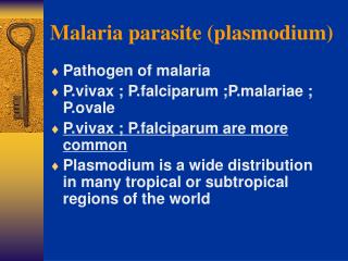
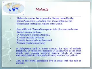
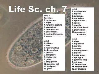
Plasmodium & Babesiosis PLASMODIA • P A R A S I T O L O G Y • DEFINITION • The plasmodia are sporozoa in which the sexual and asexual cycles of reproduction are completed in different host species. The sexual phase occurs within the gut of mosquitoes. These arthropods subsequently transmit the parasite while feeding on a vertebrate host. Within the red blood cells (RBCs) of the vertebrate, the plasmodia reproduce asexually; they eventually burst from the erythrocyte and invade other uninvolved RBCs. This event produces periodic fever and anemia in the host, a disease process known as malaria. Of the many species of plasmodia, four are known to infect humans and will be considered here: Plasmodium vivax, P. ovale, P. malariae, and P. falciparum.
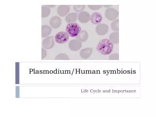
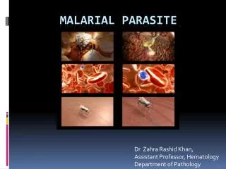
Morphology The morphology of the stained intraerythrocytic parasites shows three characteristic features aid in the identification of plasmodia: red nuclear chromatin; blue cytoplasm; and brownish-black malarial pigment, or hemozoin, consisting largely of a hemoglobin degradation product, ferriprotoporphyrin IX. The change in the shape of the cytoplasm and the division of the chromatin at different stages of parasite development are obvious. Gametocytes can be differentiated from the asexual forms by their large size and lack of nuclear division. Some of the infected erythrocytes develop membrane invaginations or caveolae-vesicle complexes, which are thought to be responsible for the appearance of the pink Schüffner’s dots or granules.
The appearance of each of the four species of plasmodia that infect humans is sufficiently different to allow their differentiation in stained smears. The parasitized erythrocyte in P. vivax and P. ovaleinfections is pale, enlarged, and contains numerous Schüffner’s dots. All asexual stages (trophozoite, schizont, merozoite) may be seen simultaneously. Cells infected by P. ovaleare elongated and frequently irregular or fimbriated in appearance. In P. malariae infections, the RBCs are not enlarged and contain no granules. The trophozoites often present as “band” forms, and the merozoites are arranged in rosettes around a clump of central pigment.
In P. falciparum infections, the rings are very small and may contain two chromatin dots rather than one. There is often more than one parasite per cell, and parasites are frequently seen lying against the margin of the cell. Intracytoplasmic granules known as Maurer’s dots may be present but are often cleft shaped and fewer in number than Schüffner’s dots. Schizonts and merozoites are not present in the peripheral blood. Gametocytes are large and banana shaped.
LIFE CYCLE OF MALARIAL PARASITES • Sporogony, or the sexual cycle, begins when a female mosquito of the genus Anopheles ingests circulating male and female gametocytes while feeding on a malarious human. In the gut of the mosquito, the gametocytes mature and effect fertilization. The resulting zygote penetrates the mosquito’s gut wall, lodges beneath the basement membrane, and vacuolates to form an oocyst. Within this structure, thousands of sporozoites are formed. The enlarging cyst eventually ruptures, releasing the sporozoites into the body cavity of the mosquito. Some penetrate the salivary glands, rendering the mosquito infectious for humans. The time required for the completion of the cycle in mosquitoes varies from 1 to weeks, depending on the species of insect and parasite as well as on the ambient temperature and humidity.
Schizogony, the asexual cycle, occurs in the human and begins when the infected Anopheles takes a blood meal from another individual. Sporozoites from the mosquito’s salivary glands are injected into the human’s subcutaneous capillaries and circulate in the peripheral blood. Within 1 hour they attach to and invade liver cells (hepatocytes). In P. vivaxand P. ovale infections, some of the sporozoites enter a dormant state immediately after cell invasion. The remaining sporozoites initiate exoerythrocytic schizogony, each producing about 2000 to 40,000 daughter cells, or merozoites. One to two weeks later, the infected hepatocytes rupture, releasing merozoites into the general circulation.
The erythrocytic phase of malaria starts with the attachment of a released hepatic merozoite to a specific receptor on the RBC surface. After attachment, the merozoite invaginates the cell membrane and is slowly endocytosed. The intracellular parasite initially appears as a ring-shaped trophozoite, which enlarges and becomes more active and irregular in outline. Within a few hours, nuclear division occurs, producing the multinucleated schizont. Cytoplasm eventually condenses around each nucleus of the schizont to form an intraerythrocytic cluster of 6 to 24 merozoite daughter cells. About 48 (P. vivax, P. ovale, and P. falciparum) to 72 (P. malariae) hours after initial invasion, infected erythrocytes rupture, releasing the merozoites and producing the first clinical manifestations of disease.
Other daughter cells are transformed into sexual forms or gametocytes. These latter forms do not produce RBC lysis, and continue to circulate in the peripheral vasculature until ingested by an appropriate mosquito. The recurring asexual cycles continue, involving an ever-increasing number of erythrocytes until finally the development of host immunity brings the erythrocytic cycle to a close. The dormant hepatic sporozoites of P. vivax and P. ovalesurvive the host’s immunologic attack, and may, after a latent period of months to years, resume intrahepatic multiplication. This leads to a second release of hepatic merozoites and the initiation of another erythrocytic cycle, a phenomenon known as relapse.
M A L A R I A • Malaria is a febrile illness caused by a parasitic infection of human erythrocytes transmitted by the bite of a mosquito. The fevers are accompanied by headache, sweats, malaise, and typically appear in paroxysmal episodes lasting hours and recurring for weeks. Complications due to capillary blockade can be fatal, particularly in the brain.
EPIDEMIOLOGY • Malaria has a worldwide distribution between 45° N and 40° S latitude, generally at altitudes below 1800 m. P. vivaxis the most widely distributed of the four species, and together with the uncommon P. malariae, is found primarily in temperate and subtropical areas. P. falciparumis the dominant organism of the tropics. P. ovaleis rare and found principally in Africa. • The intensity of malarial transmission in an endemic area depends on the density and feeding habits of suitable mosquito vectors and the prevalence of infected humans, who serve as parasite reservoirs..
In hyperendemic areas (areas where more than half of the population is parasitemic), transmission is usually constant, and disease manifestations are moderated by the development of immunity. • Mortality is largely restricted to infants and to nonimmune adults who migrate into the region. • When the prevalence of disease is lower, transmission is typically intermittent. In this situation, solid immunity does not develop and the population suffers repeated, often seasonal, epidemics, the impact of which is shared by people of all ages.
PATHOGENESIS The fever, anemia, circulatory changes, and immunopathologic phenomena characteristic of malaria are all the result of erythrocytes invasion by the plasmodia. • Fever Fever, the hallmark of malaria, appears to be initiated by the process of RBC rupture that leads to the liberation of a new generation of merozoites (sporulation). To date, all attempts to detect the factor( s) mediating the fever have been unsuccessful.
It is possible that parasite-derived pyrogens are released at the time of sporulation; alternatively, the fever might result from the release of interleukin-1 (IL-1) and/ or tumor necrosis factor (TNF) from macrophages involved in the ingestion of parasitic or erythrocytic debris. Early in malaria, RBCs appear to be infected with malarial parasites at several different stages of development, each inducing sporulation at a different time. The resulting fever is irregular and hectic. Because temperatures in excess of 40° C destroy mature parasites, a single population eventually emerges, sporulation is synchronized, and fever occurs in distinct paroxysms at 48hour or, in the case of P. malariae, 72-hour intervals. Periodicity is seldom seen in patients who are rapidly diagnosed and treated.
Anemia • Parasitized erythrocytes are phagocytosed by a stimulated reticuloendothelial system or are destroyed at the time of sporulation. At times, the anemia is disproportionate to the degree of parasitism. Depression of marrow function, sequestration of erythrocytes within the enlarging spleen, and accelerated clearance of nonparasitized cells all appear to contribute to the anemia. The mechanisms responsible for the latter are unclear. Intravascular hemolysis, although uncommon, may occur, particularly in falciparum malaria. When hemolysis is massive, hemoglobinuria develops, resulting in the production of dark urine. This process in conjunction with malaria is known as blackwater fever.
Circulatory Changes • The high fever results in significant vasodilatation. In falciparum malaria, vasodilatation leads to a decrease in the effective circulating blood volume and hypotension, which may be aggravated by other changes in the small vessels and capillaries. The intense parasitemias P. falciparum is capable of producing and the adhesion of infected RBCs to the endothelium of visceral capillaries can impair the microcirculation and precipitate tissue hypoxia, lactic acidosis, and hypoglycemia. Although all deep tissues are involved, the brain is the most intensely affected. • Excessive TNF levels might precipitate cerebral malaria by directly inducing hypoglycemia and lactic acidosis.
Other Pathogenic Phenomena • Thrombocytopenia is common in malaria and appears to be related to both splenic pooling and a shortened platelet lifespan. Both direct parasitic invasion and immune mechanisms may be responsible. There may be an acute transient glomerulonephritis in falciparum malaria and progressive renal disease in chronic P. malariae malaria. These phenomena probably result from the host immune response, with deposition of immune complexes in the glomeruli. • .
IMMUNITY • Once infected, the host quickly mounts a species- and strain-specific immunologic response that typically limits parasite multiplication and moderates the clinical manifestations of disease, without eliminating the infection-a phenomenon referred to as premunition. A prolonged recovery period marked by recurrent exacerbations in both symptoms and number of erythrocytic parasites follows. With time, these recrudescences become less severe and less frequent, eventually stopping altogether.
M A L A R I A: C L I N I C A L A S P E C T S • MANIFESTATIONS • The incubation period between the bite of the mosquito and the onset of disease is approximately 2 weeks. With P. malariae and with strains of P. vivax in temperate climates, however, this period is often more prolonged. Individuals who contract malaria while taking antimalarial suppressants may not experience illness for many months. In the United States, the interval between entry into the country and onset of disease exceeds 1 month in 25% of P. falciparum infections and 6 months in a similar proportion of P. vivax cases.
The clinical manifestations vary with the species of plasmodia but typically include chills, fever, splenomegaly, and anemia. The hallmark of disease is the malarial paroxysm. This manifestation begins with a cold stage, which persists for 20 to 60 minutes. During this time, the patient experiences continuous rigors and feels cold. With the consequent increase in body temperature, the rigors cease and vasodilatation commences, ushering in a hot stage. The temperature continues to rise for 3 to 8 hours, reaching a maximum of 40 to 41.7° C before it begins to fall. The wet stage consists of a decrease in fever and profuse sweating. It leaves the patient exhausted but otherwise well until the onset of the next paroxysm.
Typical paroxysms first appear in the second or third week of fever, when parasite sporulation becomes synchronized. In falciparum malaria, synchronization may never take place, and the fever may remain hectic and unpredictable. The first attack is often severe and may persist for weeks in the untreated patient. Eventually the paroxysms become less regular, less frequent, and less severe. Symptoms finally cease with the disappearance of the parasites from the blood.
In falciparum malaria, capillary blockage can lead to several serious complications. When the central nervous system is involved (cerebral malaria), the patient may develop delirium, convulsions, paralysis, coma, and rapid death. Acute pulmonary insufficiency frequently accompanies cerebral malaria, killing about 80% of those involved. When splanchnic capillaries are involved, the patient may experience vomiting, abdominal pain, and diarrhea with or without bloody stools. Jaundice and acute renal failure are also common in severe illness. These pernicious syndromes generally appear when the intensity of parasitemia exceeds 100,000 organisms per cubic millimeter of blood. Most deaths occur within 3 days.
DIAGNOSIS • Malarial parasites can be demonstrated in stained smears of the peripheral blood in virtually all symptomatic patients. Typically, capillary or venous blood is used to prepare both thin and thick smears, which are stained with Wright or Giemsastain and examined for the presence of erythrocytic parasites. Thick smears, in which erythrocytes are lysed with water before staining, concentrate the parasites and allow detection of very mild parasitemia. Artifacts are numerous in thick smears, and correct interpretation requires experience. The morphologic differences among the four species of plasmodia allow their speciation on the stained smear by the skilled observer.
Simple, specific card antigen detection procedures are now available. The most widely used test, ParaSightF, detects a protein (HRP2) excreted by P. falciparum within minutes. The test can be performed under field conditions and has a sensitivity more than 95%. • A second rapid test, OptiMAL, detects parasite lactate dehydrogenase, and, unlike ParaSight F, can distinguish between P. falciparum and P. vivax. • Serologic tests are offered at a few large reference laboratories but are used primarily for epidemiologic purposes. They are occasionally helpful in speciation and detection of otherwise occult infections. The recently completed sequencing of the malaria genome will lead to newer diagnostic methods.
TREATMENT • The indications for treatment rest on two factors. The first is the infecting species of Plasmodium, and the second is the immune status of the afflicted patient. Falciparum malaria is potentially lethal in nonimmune individuals such as new immigrants or travelers to a malarious area and immunosuppressed indigenous individuals such as pregnant women. These individuals must be treated emergently.
The complete treatment of malaria requires the destruction of three parasitic forms: the erythrocytic schizont, the hepatic schizont, and the erythrocytic gametocyte. The first terminates the clinical attack, the second prevents relapse, and the third renders the patient noninfectious to Anopheles and thus breaks the cycle of transmission. • Unfortunately, no single drug accomplishes all three goals.
Termination of Acute Attack • Several agents can destroy asexual erythrocytic parasites. Chloroquine, a 4-aminoquinoline, has been the most commonly used. It acts by inhibiting the degradation of hemoglobin, thereby limiting the availability of amino acids necessary for growth.
When originally introduced, it was rapidly effective against all four species of plasmodia and, in the dosage used, free of serious side effects. However, chloroquine-resistant strains of P. falciparum are now widespread in Africa and Southeast Asia, and less frequently, in other areas of Asia and in Central America and South America. • Other schizonticidal agents include quinine/ quinidine, antifolate-sulfonamide combinations, mefloquine, halofantrine, and the artemisinins.
Radical Cure • In P. vivax and P. ovale infections, hepatic schizonts persist and must be destroyed to prevent reseeding of circulating erythrocytes with consequent relapse. Primaquine, an 8-aminoquinaline, is used for this purpose. Some P. vivax infections acquired in Southeast Asia and New Guinea fail initial therapy due to relative resistance to this 8-amino-quinaline. Retreatment with a larger dose of primaquine is usually successful.
Unfortunately, primaquine may induce hemolysis in patients with G6PD deficiency. Persons of Asian, African, and Mediterranean ancestry should thus be screened for this abnormality before treatment. • Chloroquine destroys the gametocytes of P. vivax, P. ovale, and P. malariae but not those of P. falciparum. Primaquine and artemisinins, however, are effective for this latter species.
PREVENTION • Personal Protection • In endemic areas, mosquito contact can be minimized with the use of house screens, insecticide bombs within rooms, and/ or insecticide-impregnated mosquito netting around beds. Those who must be outside from dusk to dawn, the period of mosquito feeding, should apply insect repellent and wear clothing with long sleeves and pants. In addition, it is possible to suppress clinical manifestations of infection, should they occur, with a weekly dose of chloroquine.
In areas where chloroquine-resistant strains are common, an alternative schizonticidal agent should be used. Mefloquine or doxycycline are usually preferred. The antifolate pyrimethamine plus a sulfonamide can be taken as well. • General • Malaria control measures have been directed toward reducing the infected human and mosquito populations to below the critical level necessary for sustained transmission of disease. The techniques employed include those mentioned previously, treatment of febrile patients with effective antimalarial agents, chemical or physical disruption of mosquito breeding areas, and use of residual insecticide sprays.
Vaccines • Three advances in the last decade have produced the hope that an effective malaria vaccine might be within reach of medical science for the first time. The establishment of a continuous in vitro culture system provided the large quantities of parasite needed for antigenic analysis. Development of the hybridoma technique allowed the preparation of monoclonal antibodies with which antigens responsible for the induction of protective immunity could be identified. Finally, recombinant DNA procedures enabled scientists to clone and sequence the genes encoding such antigens, permitting the amino acid structure to be determined and peptide sequences suitable for vaccine development to be identified.
Babesiosis • Babesiosis is an infection of red blood cells caused by the single-celled parasite, Babesia microti, which is spread to humans by a tick bite. • Babesiosis most commonly affects domestic and wild animals and can be a serious problem in cattle. In most cases the protozoal species is specific to a single host. The organisms enter the blood via a tick bite, then infect the red blood cells where they reproduce by cell division.
Ticks are small, blood-sucking arachnids. Babesia microtiis spread to humans through the bite of the tick Ixodes scapularis(also called Ixodes dammini). Ixodes scapularis, called the "blacklegged deer tick," usually feeds on deer and mice. A tick picks up the parasites by feeding on an infected mouse and then passes them on by biting a new host, possibly a human. To pass on the parasites, the tick must be attached to the skin for 36-48 hours. Once in the bloodstream, Babesia microtienters a red blood cell, reproduces by cell division, and destroys the cell. Humans infected with Babesia microti produce antibodies that can be helpful in diagnosing the infection.
Human babesiosis, sometimes called Nantucket fever, was first diagnosed after an outbreak on Nantucket Island, off the coast of Massachusetts, in the 1970s. The causative organism, Babesia microti, is related to the one that causes malaria, and is transmitted by the deer tick that also hosts the organisms that cause Lyme disease and human erhlichiosis.
Causes and symptoms • Babesia microti live and divide within red blood cells, destroying the cells and causing anemia. The majority of people who are infected have no visible symptoms. In those who become ill, symptoms appear one to six weeks following the tick bite. Because the ticks are small, many patients have no recollection of a tick bite. The symptoms are flu-like and include tiredness, loss of appetite, fever, drenching sweats, and muscle pain. Nausea, vomiting, headache, shaking chills, blood in the urine, and depression can occur.
Diagnosis • Babesiosis is easy to diagnose but only if it is suspected. It will not show up on any routine tests. It must be suspected when a persons with exposure in an endemic area develops persistent fevers and hemolytic anemia. Babesiosis can be diagnosed by direct examination of the blood, with serology, or with PCR-based tests. Other laboratory findings include decreased numbers of red blood cells and platelets on complete blood count.
Epidemiology • Babesiosis is a vector-borne illness usually transmitted by ticks. • In babesia-endemic areas, the organism can also be transmitted by blood transfusion. • It is sometimes called "The Malaria of The North East." • While most severe cases occur in the very young, very old, or persons with underlying medical conditions (such as immunodeficiency) and those without a spleen, they can occur in normal individuals.
Treatment • Most cases of babesiosis resolve without any specific treatment. For ill patients, treatment is usually a two-drug regimen. The traditional regimen of quinine and clindamycin is often poorly tolerated; recent evidence suggests that a regimen of atovaquone and azithromycin can be equally effective. In life-threatening cases, exchange transfusion is performed. In this procedure, the infected red blood cells are removed and replaced with fresh ones.
Prevention • The only prevention for babesiosis is to minimize exposure to ticks by staying on trails when walking through the woods, avoiding tall grasses, wearing long sleeves and tucking pant legs into socks, wearing insect repellent, and checking for ticks after an outing. Remove a tick as soon as possible by grasping the tick with tweezers and gently pulling. Splenectomized people should avoid northeastern coastal regions during the tick season.