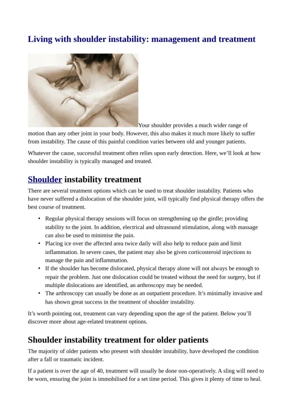
Shoulder Instability
Shoulder Instability. Dr.Syed Imran. Definition. Glenohumeral instability is the inability to maintain the humeral head in the glenoid fossa . Normal Anatomy. B ony anatomy of the shoulder joint does not provide inherent stability.


Shoulder Instability
E N D
Presentation Transcript
Shoulder Instability Dr.SyedImran
Definition • Glenohumeral instability is the inability to maintain the humeral head in the glenoidfossa.
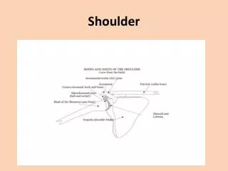
Normal Anatomy • Bony anatomy of the shoulder joint does not provide inherent stability. • Only one fourth of the large humeral head articulates with the glenoid at any given time. • The glenoid is deepened by 50% by the presence of the glenoid labrum.
Labrum increases the humeral contact to 75%. • The labrum may serve as a “chock block” to prevent excessive humeral head rollback
Superior Glenohumeral Ligament • SGHL • O = tubercle on glenoid just post to long head biceps. • I = humeral head near upper end of lesser tubercle. • Resists infsubluxation and contributes to stability in post and inf directions.
MGHL • O= sup glenoid and labrum. • I= blends with subscapularis tendon. • Limits ant excursion instability especially with arm in 45 deg abd position. • Limits ext rotation.
IGHL • O= ant glenoid rim and labrum. • I= inf aspect of humeral articular surface and anatomic neck. • 3 bands, anterior, axillary and posterior. • IGHL complex acts like a sling and when is the most important single ligamentous stabilizer in the shoulder. • Primary restraint is at 45-90 deg abd.
Pathologic Anatomy • Traumatic detachment of the glenoid labrum has been called the Bankartlesion. • Excessive laxity of the shoulder capsule. • Excessive laxity can be caused by a congenital collagen deficiency, shown by hyperlaxity of other joints, or by plastic deformation of the capsuloligamentous complex from a single macrotraumatic event or repetitive microtraumaticevents.
Hill-Sachs lesion is a defect in the posterolateral aspect of the humeral head. • Erosion of the anterior glenoid rim, stretching of the anterior capsule and subscapularis tendon, and fraying and degeneration of the glenoid labrum all can occur with repeated dislocation.
Classification • Congenital • Direction • Unidirectional • Multidirectional • Bidirectional • Degree • Subluxation / Dislocation • Duration : Acute, Subacute, Chronic, Recurrent
Type of trauma • Macrotraumatic, in which a single traumatic event results in dislocation. • Microtraumatic (acquired), in which repetitive trauma at the extremes of motion results in plastic deformation of the capsulolabral complex.
Matsen’s classification • TUBS (traumatic, unidirectional Bankart surgery) . • AMBRII (atraumatic, multidirectional, bilateral, rehabilitation, inferior capsular shift, and internal closure).
History • Physical examination • Radiographic examination • CT scan • MRI scan • Examination under Anaesthesia and arthroscopy
Radiographic examination Radiographic technique for apical oblique view of shoulder.
Radiographic technique for West Point view of shoulder to show glenoid labrum lesions. With patient prone and pillow beneath shoulder, cassette is placed superior to shoulder.
TUBS • – Conservative • Younger patient = • Higher failure rate • – Operative • Open • – 95% success rate • – Can address capsular • Laxity • Arthroscopic • – 80% success rate • – More difficult to • Address capsular laxity
AMBRI • – Conservative • Rehab shoulder girdle • Dynamic stabilizers • Rehab entire shoulder • (Improving one area accentuates other weaker areas) • – Operative • Reserved for failed • Conservative treatment and • Continued impairment • Inferior capsular shift
Surgical treatment • Anterior stability • Has a low recurrence rate. • Has a low complication rate. • Has a low reoperation rate. • Does no harm (arthritis). • Maintains motion. • Is applicable in most cases. • Allows observation of the joint. • Corrects the pathological condition. • Is not too difficult.
TUBS lesion resulting from a macrotraumatic or microtraumatic pathological condition with plastic ligamentous deformation in association with glenolabral damage, we prefer a modified Bankart procedure, open or arthroscopic, using suture anchors
Microtraumatic lesions with subtle anterior instability shown by relocation test and no significant associated labral damage, we prefer an arthroscopic capsular imbrication procedure.
Multidirectional (AMBRII) lesions are repaired with a lateral capsular shift, as described by Neer and Foster . • Attention to closure of the rotator interval is necessary to help eliminate inferior laxity at 0 degrees abduction.
Modified Bankart repair • Drill holes near the glenoid rim at approximately the 3-o'clock, 4-o'clock, and 5:30-o'clock positions, keeping the drill bit parallel to the glenoid surface . • Place Mitek G1 anchors (Mitek Surgical Products, Norwood, Mass) with 2-0 braided nonabsorbable suture in each hole with the single barb facing away from the articular surface. • Place tension on each suture to set the anchors. During this portion of the procedure, maintain the shoulder in approximately 90 degrees of abduction and 60 degrees of external rotation for throwing athletes. • Maintain the shoulder in 60 degrees abduction and 30 to 45 degrees external rotation in nonthrowing athletes and other patients
Neer capsular shift • Anterior instability is tested with the arm in external rotation and extension at various levels of abduction. • Inferior instability is tested with the arm in 0 degrees and 45 degrees of abduction. • Posterior instability is tested with the arm in internal rotation at various levels of forward elevation.
The arm in external rotation, develop a capsular flap by detaching the reinforced part of the capsule containing the inferior glenohumeral ligament from the inferior aspect of the neck of the humerus around to the posterior aspect of the neck of the humerus.
Suture the capsular flap to the stump of the subscapularis tendon and to the part of the capsule that remains on the humerus so that the capsular flap is held against the slot of raw bone. • If preferred, suture anchors can be used to secure the capsule.
Tension on the capsular flap that is selected must eliminate the inferior pouch and reduce the posterior capsular redundancy. • Suture the inferior flap first, and draw the superior flap down over it and suture it so as to cause the middle glenohumeral ligament to reinforce the capsule anteriorly and to act as a sling against inferior subluxation.
Plastic splint (A) and sling (B) used after inferior capsular shift procedure.
Multidirectional Instability • Glenohumeralsubluxation or dislocation in multiple directions. • Primary abnormality in multidirectional instability is a loose, redundant inferior pouch. • Surgery in these patients is not indicated.
Inferior capsular shift procedure. • Principle of the procedure is to detach the capsule from the neck of the humerus and shift it to the opposite side of the calcar (inferior portion of the neck of the humerus), not only to obliterate the inferior pouch and capsular redundancy on the side of the surgical approach, but also to reduce laxity on the opposite side.
Failure of surgery • Causes of Failures • Inadequate soft-tissue healing • Ligamentous laxity • Deficient capsule • Deficient subscapularis • Deficient glenoid • Engaging Hill-Sachs
Posterior instability • Conservative treatment • Operative : • Reverse Bankart’s • Reverse Putti Platt