Download
1 / 21
210 likes | 377 Vues
Molecular mechanisms of bone reconstruction in new dental implant technology Aurelian Udristioiu , Staniloiu Mihail Emergency County Hospital Targu Jiu & UCB University, Romania, E-mail: aurelianu2007@yahoo.com. Introduce

E N D
Molecular mechanisms of bone reconstruction in new dental implant technology Aurelian Udristioiu, StaniloiuMihail Emergency County Hospital TarguJiu & UCB University, Romania, E-mail: aurelianu2007@yahoo.com
Introduce • Physiological bone remodeling is a highly coordinated process responsible for bone resorption and formation and is necessary to repair damaged bone and to maintain mineral homeostasis. In addition to the traditional bone cells (osteoclasts, osteoblasts, and osteocytes) that are necessary for bone remodeling, several immune cells have also been implicated in bone disease. Periodontitis is a common disease characterized by resorption of the alveolar bone and is mediated by host responses against multiple commensal bacteria, (1). • Previous periodontitis studies have emphasized the importance of host damaging • bacteria, including Porphyromonasgingivalis (Pg), which possess high protease and immunosuppressive features (2). Colonization of such tissue-damaging bacteria in the oral cavity is enhanced by streptococci-induced biofilm formation and interactions with multiple commensals.
The mechanisms by which commensal species trigger bone resorption associated with periodontitis remain poorly understood. Alveolar bone loss is caused by osteoclast activation through RANKL (receptor activator of NF-κB ligand) signaling. Bacterial components are known to induce alveolar bone resorption by inducing the production of pro-inflammatory cytokines such as tumor necrosis factor-a (TNF-a) and interleukin (IL)-1b, which enhance receptor activator of RANK signaling by up regulation of receptor activator of nuclear factor kappa-B ligand (RANKL) and down-regulation of osteoprotegerin (OPG). • Previous studies emphasized the importance of neutrophils in the early phase of periodontitis development.
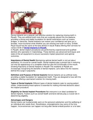
These include studies demonstrating that IL-17 affects alveolar bone resorption by inhibiting the expression of EDIL3, which prevents neutrophil recruitment at the oral bone absorption site in rodent models (3). Osteoprotegerin (OPG)2 is a soluble decoy receptor for RANKL and a physiological negative regulator of osteoclastogenesis; loss of functional OPG in mice results in animals with osteoporosis (brittle bones due to excessive osteoclastogenesis (4). • In last years, new technical of dental implants had as objective to solve rebuilding dental bone by a continue process of osteo-genesis.
The preparation samples necessary in dental inplant was made after a special manual of maxilo surgery facial, author, Anitua E, Inplantsurgerey and prosthesis. Ed. Eduardo Anitua, Puesta al Dia, Publicaciones, Vitoria, 1998. • A total of 10 patients (8 males and 2 females), between 50 and 65 years ( mean age 57.5 years) were appropriately informed that them will collected a small volume of blood, 20 cm ³, drown from peripheral vein, using sodium citrate as anticoagulant, harvesting of peripheral blood, 4.5 mL in 4 coagulation tubes of 5 ml capacity, (38% sodium citrate as an anticoagulant, 0.5 ml). • -After a short time, the 4 vacuetes of cagulation with 5 ml blood have been 8 minutes centrifugation at 4500 revs per minute (280 G), in special equipment for this tehnique.
-From each 4 tubes of plasma will be extracted from the surface of the tube, 500 microliters plasma with automatic pipette, and will release a container of wastes. • -With the same pipette from each tube, from the middle layer, extract plasma, 500 microliters plasma in biochemistry tube labeled with the number 2, (with no anticoagulant or additive, (0.5 ml x 4 = 2 ml, plasma rich in growth factors). • -With another pipette of 100 micro-liters capacity, of 3 times from each tube are aspirated the plasma layers remaining in the 4 tubes and it is delivered in biochemistry tube which is labeled with number 1. (4 x 300 microliters = 1.2 ml, layer rich in lymphocytes and megakaryocyte).
The plasma delivered in biochemistry dry vacuetts, were kipped for time a 30 minutes, in a thermostat at 37 C*, for to obtain auto-homolougous fibrin for to be used in seal at post-extraction site, to which were added proper dental bone powder filings. The patients, after 24 weeks from extraction with the alveolus sealing, made the CT scan for the evaluation of regenerated new bone density. • All patients were registered with very good results in building dental bone and followed a classic technical method of surgery maxilo-facial inplant.
Role of plasma cells and bone cells in osteogenesis of dental implant • A biotechnological alternative to accelerate alveolar bone regeneration is filling the post-exstraction alveolus with proper dental bone powder filings and preparation plasma, reach in B and T lymphocytes, monocytes, thrombocytes, megakariocytes and growth factors (PRGF). PRFG consist of small volume of platelet-rich plasma obtained and prepared rapidly and in simple manier from a patents own blood (6). In time the results provided a strong evidence for a sequential physiologic mechanism through which superficial zone(SZ) chondrocytes gain access to active transforming growth factor β (TGF-β, (7). More recently, it was reported that the reversal cells are from the osteoblast lineage, based on cell morphology, positive expression for alkaline phosphatase, and the absence of the monocyte macrophage marker MOMA-2 (monocyte + macrophage antibody-2) on these cells (8).
T-cells and B-cells • T- and B-lymphocytes are central components of the adaptive immune system that facilitate recognition and destruction of pathogens. Mice lacking either B- or T-cells have osteoporotic bones, suggesting that these immune cells participate in the maintenance of bone homeostasis during basal physiology (28). Mechanistically, mature B-cells produce >50% of total bone marrow-derived osteoprotegerin (OPG), which would contribute significantly to restraining osteoclastogenesis during normal physiology. However, the role of T-cells in regulating bone remodeling during homeostasis is less clear, (9). • Megakaryocytes • Derived from hematopoietic stem cells, megakaryocytes reside within bone marrow and produce thrombocytes (known as platelets) that are essential for normal blood clotting. In vitro, megakaryocytes enhance osteoblast proliferation and differentiation, express receptor activator of NF-κB ligand (RANKL) and OPG, and secrete an unknown soluble anti-osteoclastic factor. Overall, these data suggest that megakaryocytes have the potential to direct resorption and formation arms of bone remodeling, (10).
Osteoclasts • Osteoclasts are terminally differentiated myeloid cells that are uniquely adapted to remove mineralized bone matrix. These cells have distinct morphological and phenotypic characteristics that are routinely used to identify them, including multi-nuclearity and expression of tartrate-resistant acid phosphatase and the calcitonin receptor. CSF-1 (colony-stimulating factor 1; also known as macrophage colony-stimulating factor) and RANKL are critical cytokines required for survival, expansion, and differentiation of osteoclast precursor cells in vitro (11).Despite the current knowledge of transcription factors involved in osteoclastogenesis, the definitive physiological in vivo osteoclast precursor in mice and humans remains elusive. (12). • Osteoblasts • Osteoblasts are specialized bone-forming cells that express parathyroid hormone (PTH) receptors and have several important roles in bone remodeling: expression of osteoclastogenic factors, production of bone matrix proteins, and bone mineralization (13). In vitro, phenotypic osteoblast heterogeneity is associated with cell differentiation (14). The stage of osteoblast differentiation also influences the functional contribution of these cells to in vivo bone remodeling.
Also, osteoblasts develop from pluripotent mesenchymal stem cells that have the potential to differentiate into adipocytes, myocytes, chondrocytes, and osteoblasts under the direction of a defined suite of regulatory transcription factors. Osteoblast differentiation is controlled by the master transcription factor RUNX2 (runt-related transcription factor 2; also known as CBFA1 (core-binding factor A1)), RUNX2 null mice have a cartilaginous skeleton and completely lack mineralized tissue due to arrest of osteoblast maturation (15,16). • Disscution of Process Bone Remodeling: • Bone remodeling occurs over several weeks and is performed by clusters of bone-resorbing osteoclasts and bone-forming osteoblasts arranged within temporary anatomical structures known as “basic multicellular units” (BMUs). Traversing and encasing the BMU is a canopy of cells that creats fates a bone-remodeling compartment (17). • Daily activity places ongoing mechanical strain on the skeleton, and it is thought that osteocytes sense changes in these physical forces and translate them into biological signals that initiate bone remodeling. Damage to the bone matrix (or limb immobilization ) results in osteocyte apoptosis and increased osteoclastogenesis (18). Under basal conditions, osteocytes secrete transforming growth factor β (TGF-β), which inhibits osteoclastogenesis.
Focal osteocyte apoptosis lowers local TGF-β levels, removing the inhibitory osteoclastogenesis signals and allowing osteoclast formation to proceed (19). Following mineralization of the bone matrix, these entombed cells are called osteocytes and form a network extending throughout mineralized bone. • During bone formation, a subpopulation of osteoblasts undergoes terminal differentiation and becomes engulfed by unmineralized osteoid, at which time they are referred to as osteoid-osteocytes (20). Osteocytes are cocooned in fluid-filled cavities (lacunae) within the mineralized bone and are highly abundant, accounting for 90–95% of all bone cells (21). Osteocytes have long dendrite-like processes that extend throughout canaliculi (tunnels) within the mineralized matrix. • These dendrite-like processes interact with other osteocytes within mineralized bone and also interact with osteoblasts on the bone surface (22). Osteocytes respond to mechanical load, and this network is thought to be integral in the detection of mechanical strain and associated bone microdamage (microscopic cracks or fractures within the mineralized bone) that accumulates as a result of normal skeletal loading and fatigue (23). Data have been obtained that support the idea that osteocytes initiate and direct the subsequent remodeling process that repairs damaged bone.
Osteomacsare resident tissue macrophages that reside on or within three cells of endosteal and periosteal surfaces. In human bone, osteomacs can be identified by expression of the myeloid marker CD68, their distinctive stellate morphology, and location in close proximity to bone surfaces (24). In vitro, osteomacs are required for full functional differentiation, including mineralization, of osteoblasts, [Figure 1],(25). • The calciotropic hormone PTH is an endocrine remodeling signal generated to maintain calcium homeostasis. PTH is secreted by the parathyroid glands in response to reduced serum calcium and acts peripherally on kidneys and bone and indirectly on the intestine to maintain serum calcium homeostasis. In the bone microenvironment, PTH activates a seven-transmembrane G-protein-coupled receptor, the PTH receptor, on the surface of osteoblastic cells (26). Binding of PTH to its receptor activates protein kinase A, protein kinase C, and calcium intracellular signaling pathways in these cells (27) and induces a wave of transcriptional responses that produce/modulate secretion of molecules that recruit osteocl.ast precursors, induce osteoclast differentiation and activation, and establish bone resorption.
Schematic representation BMU and the associated bone-remodeling process. Accumulation of exogenous activated TGF-β in the superficial zone of articular cartilage build dental bone.
Conclusions • Regenerative therapies such as guided tissue regeneration (GTR) and guided bone regeneration (GBR) are universally accepted as surgical treatments not only for the repair and reconstruction of degraded periodontal tissues but also for quantitative and qualitative enhancement of host bones in localized defects of the alveolar bone.[28] • One of the best sources of growth factors in the body is blood platelets. Growth factors such as platelet-derived growth factor (PDGF) and transforming growth factor-β (TGF-β), contained in the α-granules of platelets and released at sites of injury, have been shown to be important in the normal healing of bone, gingiva, and skin.[29] • The growing application of dental implants to replace lost teeth coupled with the demand by patients to shorten the healing period following implant installation have also led to vast efforts by researchers to improve the quality of biomaterials and to develop dental implant surfaces with improved macroscopic and microscopic structures that allow enhanced osseointegration, (30). • Moreover, some researchers have tried to enhance osteogeneration rate in the peri-implant bones by employing biological factors, especially PDGFs which are typically delivered as PRP or plasma rich in growth factor (PRGF), (31,32).