Download
1 / 56
560 likes | 692 Vues
Join us for a seminar on aneuploidy and genome evolution by Dr. Andreas Madlung from the University of Puget Sound. Learn about the fascinating role of DNA polymerase I in E. coli, including its enzymatic activities and the crucial mechanics of DNA replication. Discover the structural complexities of E. coli DNA polymerase and its associated proteins, alongside visuals and models detailing the replication process. Don't miss this opportunity on April 8, 2009, at 4 PM in BI 234. Office hours for further discussion are available next week.

E N D
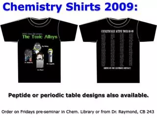
Chemistry Shirts 2009: Peptide or periodic table designs also available. Order on Fridays pre-seminar in Chem. Library or from Dr. Raymond, CB 243
“A role for aneuploidy in genome evolution?”Andreas Madlung, Associate Professor, Biology Dept., University of Puget Sound, Tacoma, WAWednesday, April 8, 2009 at 4 pm in BI 234
Chemistry Seminar F 4/10 3:15 pm SL 130 Do try to attend. This guy is good!
Office Hours next week: M 12-1 R 2-3
Figure 30-7 Priming of DNA synthesis by short RNA segments. Page 1139
DNA Polymerase I (pol I) from E. coli first DNA polymerase characterized. approximately 400 molecules of the enzyme per cell. large protein with a molecular weight of approximately 103 kDa (103,000 grams per mole). a divalent cation (Mg++) for activity Three enzymatic activities: 1. 5'-to-3' DNA Polymerase activity2. 3'-to-5' exonuclease (Proofreading activity)3. 5'-to-3' exonuclease (Nick translation activity) It is possible to remove the 5'-to-3' exonuclease activity using a protease to cut DNA pol I into two protein fragments Both the polymerization and 3'-to-5' exonuclease activities are on the large Klenow fragment of DNA pol I, and the 5'-to-3' exonuclease activity is on the small fragment.
Like all known DNA polymerases, DNA polymerase I requires a primer from which to initiate replication and polymerizes deoxyribonucleotides into DNA in the 5' to 3' direction using the complementary strand as a template. Newly synthesized DNA is covalently attached to the primer, but only hydrogen-bonded to the template. The template provides the specificity according to Watson-Crick base pairing 4. Only the alpha phosphate of the dNTP is incorporated into newly synthesized DNA
Figure 30-8b X-Ray structure of E. coli DNA polymerase I Klenow fragment (KF) in complex with a dsDNA (a tube-and-arrow representation of the complex in the same orientation as Part a). Page 1141
Figure 30-12 Nick translation as catalyzed by Pol I. Page 1144
Figure 30-8a X-Ray structure of E. coli DNA polymerase I Klenow fragment (KF) in complex with a dsDNA. Page 1141
Here’s a computer modelhttp://www.youtube.com/watch?v=4jtmOZaIvS0 Overview of DNA and replication http://207.207.4.198/pub/flash/24/menu.swf This is a pretty good outline: http://www.youtube.com/watch?v=teV62zrm2P0&NR=1 Another one with review questions http://www.wiley.com/college/pratt/0471393878/student/animations/dna_replication/index.html
Figure 30-13a X-Ray structure of the subunit of E. coli Pol III holo-enzyme. Ribbon drawing. Page 1146
Figure 30-13b The subunit of E. coli Pol III holoenzyme. Space-filling model of sliding clamp in hypothetical complex with B-DNA. Page 1146
Sliding clamp http://www.callutheran.edu/Academic_Programs/Departments/BioDev/omm/poliiib_2/poliiib.htm
Clamp loading: ·All clamp loaders utilize the energy of ATP to assemble their respective clamps onto replication forks ·Various studies have suggested that the clamp loading complex starts off in a closed form and, upon bind ATP, is drven into an open conformation that binds the clamp ( dimer) One formed, this complex between the clamp loader and the clamp binds to the DNA, inserts the DNA through the open clamp and then hydrolyzes ATP
Figure 30-14 Unwinding of DNA by the combined action of DnaB and SSB proteins.
Figure 30-15 Electron microscopy–based image reconstruction of T7 gene 4 helicase/primase. Page 1147
Figure 30-17 Active, rolling mechanism for DNA unwinding by Rep helicase.
Figure 30-19 X-Ray structure of the N-terminal 135 residues of E. coli SSB in complex with dC(pC)34. Page 1149
Figure 30-20 The reactions catalyzed by E. coli DNA ligase. Page 1150
Figure 30-21 X-Ray structure of DNA ligase from Thermus filiformis. Page 1151
Figure 31-1 The induction kinetics of b-galactosidase in E. coli. Page 1217
Total RNA Pulse-chase MVA Fig. 26.3
3 Major Types of RNA: messenger transfer ribosomal Other small RNAs are involved in splicing.
(prok) Inh. initiation Euk inhibitor Blocks elongation MVA Fig. 26.4
Table 31-1 Components of E. coli RNA Polymerase Holoenzyme. Page 1221
RNAP IV!! Table 31-2 RNA Polymerase Subunitsa. Page 1232
Figure 31-11aX-Ray structure of Taq RNAP core enzyme. a subunits are yellow and green, b subunit is cyan, b¢ subunit is pink, w subunit is gray. What do you notice about this structure? Page 1224
Figure 31-13bModel of the open (Rpo) complex of Taq RNAP with promoter-containing DNA showing the transcription bubble and the active site. Page 1225
Figure 31-14 The two possible modes of RNA chain growth. Growth may occur (a) by the addition of nucleotides to the 3¢ end and (b) by the addition of nucleotides to the 5¢ end. Page 1226 How could you distinguish between these two possibilities?
DNA Footprinting http://users.rcn.com/jkimball.ma.ultranet/BiologyPages/F/ Footprinting.html
Figure 31-10 The sense (nontemplate) strand sequences of selected E. coli promoters. Page 1223
Figure 31-12a The sequence of a fork-junction promoter DNA fragment. Numbers are relative to the transcription start site, +1. Page 1225
Figure 31-15 RNA chain elongation by RNA polymerase. Page 1227