Download
1 / 46
470 likes | 811 Vues
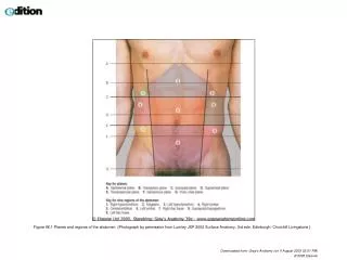
Figure 66.1 Planes and regions of the abdomen. (Photograph by permission from Lumley JSP 2002 Surface Anatomy, 3rd edn. Edinburgh: Churchill Livingstone.). Downloaded from: Gray's Anatomy (on 4 August 2005 03:01 PM). © 2005 Elsevier .

E N D
Figure 66.1 Planes and regions of the abdomen. (Photograph by permission from Lumley JSP 2002 Surface Anatomy, 3rd edn. Edinburgh: Churchill Livingstone.) Downloaded from: Gray's Anatomy (on 4 August 2005 03:01 PM) © 2005 Elsevier
Figure 66.2 Anterior abdominal wall landmarks. (Photograph by permission from Lumley JSP 2002 Surface Anatomy, 3rd edn. Edinburgh: Churchill Livingstone.) Downloaded from: Gray's Anatomy (on 4 August 2005 03:01 PM) © 2005 Elsevier
Figure 66.3 Intra-abdominal visceral landmarks. (Photograph by permission from Lumley JSP 2002 Surface Anatomy, 3rd edn. Edinburgh: Churchill Livingstone.) Downloaded from: Gray's Anatomy (on 4 August 2005 03:01 PM) © 2005 Elsevier
Figure 66.4 Retroperitoneal visceral landmarks. (Photograph by permission from Lumley JSP 2002 Surface Anatomy, 3rd edn. Edinburgh: Churchill Livingstone.) Downloaded from: Gray's Anatomy (on 4 August 2005 03:01 PM) © 2005 Elsevier
Figure 44.4 Contents of the vertebral canal in the lumbosacral region. Adapted from Mackintosh RR 1951 Lumbar Puncture and Spinal Analgesia. Edinburgh: E&S Livingstone. (Modified with permission from Mackintosh RR 1951 Lumbar Puncture and Spinal Analgesia. Edinburgh: E&S Livingstone.) Downloaded from: Gray's Anatomy (on 4 August 2005 02:12 PM) © 2005 Elsevier
Figure 44.5 The lumbar interlaminar window in extension and flexion. Downloaded from: Gray's Anatomy (on 4 August 2005 02:12 PM) © 2005 Elsevier
Figure 44.6 The position of the needle in lumbar puncture. Downloaded from: Gray's Anatomy (on 4 August 2005 02:12 PM) © 2005 Elsevier
Figure 44.7 Palpation of the sacral cornua for caudal epidural injection. With permission from Ellis H, Feldman SA 1997 Anatomy for Anaesthetists, 7th edn. Oxford: Blackwell Science. (By permission from Ellis H, Feldman S 1997 Anatomy for Anaesthetists, 7th edn. Oxford: Blackwell Science.) Downloaded from: Gray's Anatomy (on 4 August 2005 02:12 PM) © 2005 Elsevier
Figure 44.8 Position of the needle in caudal epidural injection. Downloaded from: Gray's Anatomy (on 4 August 2005 02:12 PM) © 2005 Elsevier
MORFOFISIOLOGIA MIEMBRO SUPERIOR PROF. CRISTINA ABREU
ANATOMIA REGIONAL HOMBRO • HUESOS Y ARTICULACIONES PRINCIPALES • CINTURA ESCAPULAR • - OMOPLATO (ESCAPULA) - CLAVICULA - ART. ESTERNOCLAVICULAR - ART. ACROMIOCLAVICULAR
BRAZO • HUMERO • ARTICULACION CON CAVIDAD GLENOIDEA DEL OMOPLATO.
Figure 48.5 Overview of superficial veins of the left upper limb. Downloaded from: Gray's Anatomy (on 4 August 2005 02:55 PM) © 2005 Elsevier
Figure 48.6 Lymph nodes of the left upper limb. Downloaded from: Gray's Anatomy (on 4 August 2005 02:55 PM) © 2005 Elsevier
Figure 48.13 Posterior (A) and anterior (B) views of the shoulder region. (By permission from Lumley JSP 2002 Surface Anatomy, 3rd edn. Edinburgh: Churchill Livingstone.) Downloaded from: Gray's Anatomy (on 4 August 2005 02:55 PM) © 2005 Elsevier
Figure 48.14 Anterior view of the arm abducted at the shoulder. (Photograph by Sarah-Jane Smith.) Downloaded from: Gray's Anatomy (on 4 August 2005 02:55 PM) © 2005 Elsevier
Figure 48.15 Posterior view of the arm abducted at the shoulder. (Photograph by Sarah-Jane Smith.) Downloaded from: Gray's Anatomy (on 4 August 2005 02:55 PM) © 2005 Elsevier
Figure 48.16 Volar surface of the distal forearm and hand. (Photograph by Sarah-Jane Smith.) Downloaded from: Gray's Anatomy (on 4 August 2005 02:55 PM) © 2005 Elsevier
Figure 48.17 Dorsal surface of the distal forearm and hand. (Photograph by Sarah-Jane Smith.) Downloaded from: Gray's Anatomy (on 4 August 2005 02:55 PM) © 2005 Elsevier
Figure 48.18 Radial aspect of the distal forearm and wrist to show the anatomical snuffbox. (By permission from Lumley JSP 2002 Surface Anatomy, 3rd edn. Edinburgh: Churchill Livingstone.) Downloaded from: Gray's Anatomy (on 4 August 2005 02:55 PM) © 2005 Elsevier
Figure 56.1 Frontal view of trunk demonstrating bony and soft tissue structures. (By permission from Lumley JSP 2002 Surface Anatomy, 3rd edn. Edinburgh: Churchill Livingstone.) Downloaded from: Gray's Anatomy (on 4 August 2005 02:55 PM) © 2005 Elsevier
Figure 56.2 Posterior aspect of the trunk to show surface anatomy, bony and soft tissue structures. (By permission from Lumley JSP 2002 Surface Anatomy, 3rd edn. Edinburgh: Churchill Livingstone.) Downloaded from: Gray's Anatomy (on 4 August 2005 02:55 PM) © 2005 Elsevier
Figure 56.3 Lateral view of trunk demonstrating bony and soft tissue structures. (Photograph by Sarah-Jane Smith.) Downloaded from: Gray's Anatomy (on 4 August 2005 02:55 PM) © 2005 Elsevier
Figure 56.4 Frontal view of trunk demonstrating the surface anatomy of the heart and optimal sites for auscultation. Downloaded from: Gray's Anatomy (on 4 August 2005 02:55 PM) © 2005 Elsevier
Figure 44.1 Back view of trunk. (Photograph by Sarah-Jane Smith.) Downloaded from: Gray's Anatomy (on 4 August 2005 02:12 PM) © 2005 Elsevier
Figure 44.2 Back view of trunk, arms abducted. (Photograph by Sarah-Jane Smith.) Downloaded from: Gray's Anatomy (on 4 August 2005 02:12 PM) © 2005 Elsevier
Figure 44.3 Back of trunk, oblique view. (Photograph by Sarah-Jane Smith.) Downloaded from: Gray's Anatomy (on 4 August 2005 02:12 PM) © 2005 Elsevier
Figure 110.12 Inguinal region (bones and soft tissues) and femoral triangle (vessels and nerves). (Photograph by Sarah-Jane Smith. Artwork modified from Lumley JSP 2002 Surface Anatomy, 3rd edn. Edinburgh: Churchill Livingstone.) Downloaded from: Gray's Anatomy (on 4 August 2005 03:10 PM) © 2005 Elsevier
Figure 110.13 Lateral aspect of the hip joint: bones. (Photograph by Sarah-Jane Smith. Artwork modified from Lumley JSP 2002 Surface Anatomy, 3rd edn. Edinburgh: Churchill Livingstone.) Downloaded from: Gray's Anatomy (on 4 August 2005 03:10 PM) © 2005 Elsevier
Figure 110.14 Gluteal region and posterior aspect of the thigh: bones. (Photograph by Sarah-Jane Smith. Artwork modified from Lumley JSP 2002 Surface Anatomy, 3rd edn. Edinburgh: Churchill Livingstone.) Downloaded from: Gray's Anatomy (on 4 August 2005 03:10 PM) © 2005 Elsevier
Figure 110.16 Medial aspect of the flexed knee: bone and muscles. (Photograph by Sarah-Jane Smith. Artwork modified from Lumley JSP 2002 Surface Anatomy, rd edn. Edinburgh: Churchill Livingstone.) Downloaded from: Gray's Anatomy (on 4 August 2005 03:11 PM) © 2005 Elsevier
Figure 110.15 Anterior and medial aspect of the thigh: bones. (Photograph by Sarah-Jane Smith. Artwork modified from Lumley JSP 2002 Surface Anatomy, 3rd edn. Edinburgh: Churchill Livingstone.) Downloaded from: Gray's Anatomy (on 4 August 2005 03:10 PM) © 2005 Elsevier
Figure 110.17 Lateral aspect of the flexed knee: bone and soft tissues. (Photograph by Sarah-Jane Smith. Artwork modified from Lumley JSP 2002 Surface Anatomy, 3rd edn. Edinburgh: Churchill Livingstone.) Downloaded from: Gray's Anatomy (on 4 August 2005 03:11 PM) © 2005 Elsevier
Figure 110.18 Anterior aspect of the lower leg: bones. (Photograph by Sarah-Jane Smith. Artwork modified from Lumley JSP 2002 Surface Anatomy, 3rd edn. Edinburgh: Churchill Livingstone.) Downloaded from: Gray's Anatomy (on 4 August 2005 03:11 PM) © 2005 Elsevier
Figure 110.19 Left foot and ankle, lateral view. (Photograph by Sarah-Jane Smith.) Downloaded from: Gray's Anatomy (on 4 August 2005 03:11 PM) © 2005 Elsevier
Figure 110.20 Sole of the foot: bones. (Photograph by Sarah-Jane Smith. Artwork modified from Lumley JSP 2002 Surface Anatomy, 3rd edn. Edinburgh: Churchill Livingstone.) Downloaded from: Gray's Anatomy (on 4 August 2005 03:11 PM) © 2005 Elsevier
Figure 110.21 Gluteal region and posterior aspect of the thigh: superficial (left limb) and deep (right limb) muscles. (Photograph by Sarah-Jane Smith. Artwork modified from Lumley JSP 2002 Surface Anatomy, 3rd edn. Edinburgh: Churchill Livingstone.) Downloaded from: Gray's Anatomy (on 4 August 2005 03:11 PM) © 2005 Elsevier
Figure 110.22 Anterior and medial aspect of the thigh: superficial (right limb) and deep (left limb) muscles. (Photograph by Sarah-Jane Smith. Artwork modified from Lumley JSP 2002 Surface Anatomy, 3rd edn. Edinburgh: Churchill Livingstone.) Downloaded from: Gray's Anatomy (on 4 August 2005 03:11 PM) © 2005 Elsevier
Figure 110.23 Popliteal fossa: soft tissues. (By permission from Lumley JSP 2002 Surface Anatomy, 3rd edn. Edinburgh: Churchill Livingstone.) Downloaded from: Gray's Anatomy (on 4 August 2005 03:11 PM) © 2005 Elsevier
Figure 110.24 Anterior aspect of the lower leg: muscles. (Photograph by Sarah-Jane Smith. Artwork modified from Lumley JSP 2002 Surface Anatomy, 3rd edn. Edinburgh: Churchill Livingstone.) Downloaded from: Gray's Anatomy (on 4 August 2005 03:11 PM) © 2005 Elsevier
Figure 110.26 Left leg and foot, lateral view with ankle dorsiflexed. (Photograph by Sarah-Jane Smith.) Downloaded from: Gray's Anatomy (on 4 August 2005 03:11 PM) © 2005 Elsevier
Figure 110.27 Left leg and foot, ankle plantar flexed, lateral view. (Photograph by Sarah-Jane Smith.) Downloaded from: Gray's Anatomy (on 4 August 2005 03:11 PM) © 2005 Elsevier
Figure 110.28 Surface markings of the sciatic nerve. The line of the nerve joins the midpoint between the ischial tuberosity and the posterior superior iliac spine with the midpoint between the ischial tuberosity and the greater trochanter and then continues vertically down the back of the thigh. (From Ellis H, Feldman S 1997 Anatomy for Anaesthetists, 7th edn. Oxford: Blackwell Science. By permission of Blackwell Publishing.) Downloaded from: Gray's Anatomy (on 4 August 2005 03:11 PM) © 2005 Elsevier
Figure 110.25 Left calf and ankle, posterior view with foot plantigrade. (Photograph by Sarah-Jane Smith.) Downloaded from: Gray's Anatomy (on 4 August 2005 03:11 PM) © 2005 Elsevier
“La educación no se trata de cuanto tienes alojado en la memoria,o siquiera cuanto sabes. Es saber diferenciar lo que sabes de lo que no sabes” Anatole France