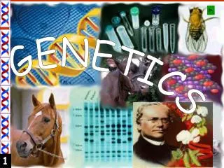
Understanding Chromosomes and Sex Determination in Humans
This article explores the structure and function of chromosomes, focusing on the mechanisms of sex determination in humans. Chromosomes, composed of DNA and proteins, come in paired sets, with each individual having 46 chromosomes organized into 23 pairs, including two sex chromosomes. The unique roles of X and Y chromosomes in determining male and female characteristics are examined. Additionally, the process of meiosis is described, highlighting how gametes are produced to ensure genetic diversity while maintaining chromosome numbers across generations.

Understanding Chromosomes and Sex Determination in Humans
E N D
Presentation Transcript
Genetics Topic 4 and 10
Why sex???? • http://www.pbs.org/wgbh/nova/miracle/program.html
Chromosomes • Chromosomes are composed of DNA and protein (histone) • Each body cell, called a somatic cell, is composed of 46 chromosomes (23 pairs). • Each chromosome consists of two sister chromatids joined at the centromere.
Chromosomes • Two chromosomes composing a pair are called homologous chromosomes (or homologues) because they both carry genes controlling the same inherited characteristics. • For example, if a gene that determines whether a person has freckles is located at a particular place, or locus, then the other chromosome of the homologous pair also has a gene for freckles at that locus • However, the two homologues may have different variations of the freckles genes (called alleles), perhaps one that promotes freckles and one that does not.
Chromosomes • Sex chromosome • X and Y chromosomes • Determine the sex of an individual, and carry genes that perform other functions as well. • 23rd pair of chromosomes in humans • The two distinct chromosomes X and Y are important exception to the general pattern of homologous chromosomes. • Human females have a homologous pair of X chromosomes (XX), but males have one X and one Y chromosome (XY). • Only small parts of the X and Y are homologous; most of the genes carried on the X chromosome do not have counterparts on the tiny Y, and the Y chromosome has genes lacking on the X.
Chromosomes • The other 22 pairs of chromosomes are autosomes, or “body chromosomes”. • For both autosomes and sex chromosomes, we inherit one chromosome of each pair form our mother and other from our father. *a key factor in the human life cycle and in the life cycles of all other species that reproduce sexually.
Chromosomes • Any cell with two homologous sets of chromosomes is called a diploid cell, and the total number of chromosomes is called the diploid number (abbreviated 2n) • For humans, the diploid number is 46; that is 2n=46 • Almost all human cells are diploid
Chromosomes • The exception are the egg and sperm cells, collectively known as gametes. • Each gamete has a single set of chromosomes: 22 autosomes plus a single sex chromosome,either X or Y. • A cell with a single chromosome set is called a haploid cell. • For humans, the haploid number (abbreviated n) is 23; that is n=23
Chromosomes • In humans, sexual intercourse allows a haploid sperm cell from the father to reach and fuse with a haploid egg cell of the mother in the process of fertilization. • The resulting fertilized egg is called a zygote, which is now diploid. • It has two haploid sets of chromosomes: one set from the mother and a homologous set from the father. • The life cycle is completed as a sexually mature adult develops from the zygote. • Mitotic cell division ensures that all somatic cells of the human body receive copies of all of the zygote’s 46 chromosomes.
Chromosomes • All sexual life cycles involve an alternation of diploid and haploid stages. • Having haploid gametes keeps the chromosome number from doubling in each generation. • Gametes are made by a special sort of cell division called meiosis, which occurs only in reproductive organs (ovaries and testes) • Whereas mitosis produces daughter cells with the same numbers of chromosomes as the parent cell, meiosis reduces the chromosome number in half.
Meiosis • Meiosis • Type of cell division that produces haploid gametes in diploid organisms. • Many of the stages of meiosis closely resemble corresponding stages in mitosis. • Meiosis, like mitosis, is preceded by the replication of chromosomes. • However, this single replication is followed by two consecutive cell divisions, called Meiosis I and Meiosis II. • These divisions result in four daughter cells, each with a single haploid set of chromosomes. • Thus, meiosis produces daughter cells with only half as many chromosomes as the parent cell.
Interphase • Like mitosis, meiosis begins by interphase, during which the chromosomes duplicate. • Occurs when the cell is between cell division • Interphase stages: • G1: Cells grow to mature size • S: DNA is copied • G2: Cell prepares for division • At the end of interphase, each chromosomes consists of two genetically identical sister chromatids attached together. • Chromosomes are not yet visible under the microscope; they are in a form called chromatin
Prophase I • Most complex phase of meiosis and typically occupies over 90% of the time required for meiotic cell division. • Chromatin coils up so that individual chromosomes become visible • A process called synapsis occurs, and homologous chromosomes, each composed of two sister chromatids, come together as pairs. • Resulting structure, consisting of four chromatids, is called a tetrad. • Chromatids of homologous chromosomes exchange segments in a process called crossing over: • Rearranges genetic information, since homologues may be different from each other. • This genetic shuffling makes an important contribution to the genetic variability resulting from sexual reproduction.
Prophase I • As prophase I continues, the chromosomes condense further as the nucleoli disappear. • Spindle fiber forms • Nuclear envelope breaks down
Metaphase I • The chromosome tetrads are aligned on the metaphase plate, midway between the two poles of the spindle. • Each chromosome is condensed and thick, with its sister chromatids still attached at their centromeres • Spindle microtubules are attached at centromeres. • In each tetrad, the homologous chromosomes are held together at sites of crossing over. • Within each tetrad, the spindle microtubules attached to one of the homologous chromosomes from one pole of the cell, and the microtubules attached to the other homologous chrome come from the opposite pole • Getting set up to separate the homologous chromsomes!
Anaphase I • Sister chromatids remain attached, however, the tetrads split up . • The sister chromatids move to opposite poles • The cells are now containing half of the genetic information from the original parent cell and are thus considered HAPLOID!
Telophase I and Cytokinesis • Chromosomes arrive at the poles of the cell • Each pole of the cell has a haploid chromosome set, although each chromosome is still in duplicate form at this point= each chromosome still consists of two sister chromatids. • Cytokinesis occurs along with telophase I and two haploid daughter cells are formed.
Before Meiosis II… • In some organisms, the chromosomes uncoil and the nuclear envelope re-forms, and there is an interphase before meiosis II begins. • IN other species, daughter cells produced during the first meiotic division immediately begin preparation for the second meiotic division. • In either case, NO chromosome duplication occurs between telophase I and the onset of meiosis II.
Meiosis II • In organisms having an interphase after meiosis I, the chromosomes condense again and the nuclear envelope breaks down during prophase II. • In any case, meiosis II is essentially the same as mitosis. • The key difference is that meiosis II starts with a haploid cell.
Prophase II • A spindle forms and moves the chromosomes toward the middle of the cell.
Metaphase II • Chromosomes are aligned on the metaphase plate as they are in mitosis, with the centromeres of the sister chromatids pointing towards opposite poles.
Anaphase II • centromeres of sister chromatids finally separate • the sister chromatids of each pair, now individual chromosomes, move toward opposite poles of the cell.
Telophase II • Nuclei form at the cell poles, and cytokinesis occurs at the same time. • There are now four daughter cells, each with the haploid number of (single) chromosomes.
Meiosis Animation • http://www.sumanasinc.com/webcontent/animations/content/meiosis.html
Mitosis vs. Meiosis • Mitosis • Provides growth, tissue repair, and asexual reproduction • Produces daughter cells genetically identical to the parent cell • Involves one division of the nucleus, and is usually accompanied by cytokinesis, producing two diploid daughter cells. • Meiosis • Need for sexual reproduction • Entails two nuclear and cytoplasmic divisions • Yields four haploid daughter cells, with one member of each homologous chromosome pair. • Form tetrads; crossing over occurs.
Meiosis: Genetic Variation • We’ve discussed how mutations lead to genetic variation. • Also, The arrangement of homologous chromosomes pairs at metaphase of meiosis I affects the resulting gametes. • The orientation of homologous pairs in the center of the cell is random; thus producing gametes with random chromosomes • For any species, the total number of combinations of chromosomes that meiosis can package into gametes is 2n, where n is the haploid number. • For a human, 223, or about 8 million possible chromosome combinations • This means that each gamete you produce contains one of roughly 8 million possible combinations of chromosomes inherited from your mother and father.
Meiosis: Genetic Variation • Possibility when a gamete from one individual unites with a gamete from another individual in fertilization: • In humans, the random fusion of a single sperm wit a single ovum during fertilization will produce a zygote with any of 64 trillion (8 million x 8 million) combinations of chromosomes!
Meiosis: Genetic Variation • Homologous chromosomes • Bear two different kinds of genetic information for the same characterisitic • The key to what really makes gametes—and therefore offspring--different
Meiosis: Genetic Variation • Crossing Over: • An exchange of corresponding segments between two homologous chromosomes. • Occurs during prophase I of meiosis. • Chromosomes are a tetrad—four chromatids, with each pair of sister chromatids joined at their centromeres. • Each gene on each homologue is aligned precisely with the corresponding gene on the other homologue • Sites of crossing over appear as X-shaped regions; each is called a chiasma. • place where two homologous chromatids are attached to each other. • Can produce new combinations of genes= genetic recombination!
Meiosis: Genetic Variation • Crossing over: • 1. The DNA molecules of two nonsister chromatids—one maternal and one paternal—break at the same place. • 2. Immediately, the two broken chromatids join together in a new way . • In effect, the two homologous segments trade places, or cross over, producing hybrid chromosomes with new combinations of maternal and paternal genes. • Called “recombinants” • 3. When the homologous chromosomes separate in anaphase I, each contains a new segment originating form its homologues. • 4. Finally, in meiosis II, the sister chromatids separate, each going to a different gamete. **In meiosis in humans, an average of one to three crossover events occur per chromosome pair.
Meiosis: Genetic Variation • In summary, there are three sources of genetic variability, besides mutations, in sexually reproducing organisms: • 1. crossing over during prophase I • 2.independent orientation of chromosomes at metaphase 1 • 3.random fertilization
Karyotypes • The term karyotype refers to the chromosome complement of a cell or a whole organism. • A karyotype is an ordered display of magnified images of an individual’s chromosomes arranged in pairs, starting with the longest. • In particular, it shows the number, size, and shape of the chromosomes as seen during metaphase of mitosis. • Chromosome numbers vary considerably among organisms and may differ between closely related species.
Karyotypes • Karyotypes are prepared from the nuclei of cultured white blood cells that are ‘frozen’ at the metaphase stage of mitosis. • Shows the chromosomes condensed and doubled • A photograph of the chromosomes is then cut up and the chromosomes are rearranged on a grid so that the homologous pairs are placed together. • Homologous pairs are identified by their general shape, length, and the pattern of banding produced by a special staining technique.
Karyotypes • Male karyotype • Has 44 autosomes, a single X chromosome, and a Y chromosome (written as 44 + XY) • Female karyotype • Shows two X chromosomes (written as 44 + XX)
Down Syndrome • Trisomy 21; named after John Langdon Down, who characterized the syndrome in 1866 • 47 chromosomes total; there are three number 21 chromsomes • In most cases, a human embryo with an abnormal number of chromosomes is spontaneously aborted (miscarried) long before birth. • Some chromosome abnormalities upset the genetic balance less drastically, and individuals carrying them can survive.
Down Syndrome • Trisomy 21 is the most common chromosome number abnormality. • Affects about one out of every 700 children born, and is the most common serious birth defect in the US • Symptoms include characteristic • facial features, notably a round face, a skin fold at the inner corner of the eye, a flattened nose bridge • small, irregular teeth, as well as short stature • heart defects, and susceptibility to respiratory infections, leukemia, and Alzheimer’s disease. • Exhibit varying degrees of mental retardation. • Some live to middle age or beyond, and many are socially adept and able to hold jobs. • Most are sexually underdeveloped and sterile • A few women have had children, however, half of their eggs will have the extra chromosome 21, so there is a 50% chance that she will transmit the syndrome to her child.
Down Syndrome • Down Syndrome incidence: • The incidence of Down Syndrome in the offspring of normal parents increases remarkedly with the age of the mother. • Strikes less than 0.05% of children (fewer than one in 2,000) born to women under age 30. • Risk climbs to 1.25% for mothers in their early 30s and is even higher for older mothers. • Because of this relatively high risk, pregnant women over 35 are candidates for fetal testing for trisomy 21 and other chromosomal abnormalities.
Errors in Meiosis • Nondisjunction • Members of a chromosome fail to separate. • Can lead to an abnormal chromosome number in any sexually reproducing diploid organism. • For example, if there is nondisjunction affecting human chromosome 21 during meiosis I, half the resulting gametes will carry an extra chromosome 21. • Then, if one of these gametes unites with a normal gamete, trisomy 21 (Down Syndrome) will result.
Errors is Meiosis • Two ways that nondisjunction can occur: • 1.a pair of homologous chromosomes does not separate during Meiosis I. • Two of the gametes produced will n + 1(abnormal), and two will be n- 1 (abnormal). • 2. meiosis I is normal, but one pair of sister chromatids fails to separate during meiosis II. • Two of the resulting gametes are n + 1 and n-1 (abnormal) and two will be n (normal) • Abnormal gametes that get fertilized, will result in a zygote with an extra chromosome. • Mitosis will then transmit the anomaly to all embryonic cells, causing some syndrome linked to abnormal genes.
Errors in Meiosis • What causes nondisjunction? • We do not yet know the answer, nor do we fully understand why offspring with trisomy 21 are more likely to by born as a woman ages. • We do know, however, that meiosis begins in a woman’s ovaries before she is born but is not completed until years later, at the time of an ovulation. • Because only one egg usually matures each month, a cell might remain arrested in the mid-meiosis state for decades. • Some research points to an age-dependent error in one of the checkpoints that coordinates the process of meiosis.
Errors in Meiosis • Nondisjunction can also occur in sex chromosomes. • For example, Klinefelter’s Syndrome : • males have an extra X chromosome, making him XXY • occurs approximately 1 out of every 2,000 live births • Have male sex organs, but the testes are abnormally small and the individual is sterile. • Often includes breast enlargement and other female body characteristics. • Person is usually of normal intelligence