Download
1 / 34
350 likes | 467 Vues
This overview of neonatal seizures presents key insights into their pathophysiology, causes, and consequences. It highlights abnormal neuronal activities, imbalances in excitatory and inhibitory neurotransmitters, and biochemical changes during seizures. The incidence is notably higher in neonates, especially within the first ten days of life. Long-term impacts on brain development and cognition are discussed, with evidence linking prolonged seizures to lasting deficits. Understanding these factors is crucial for effective management and intervention in affected infants.

E N D
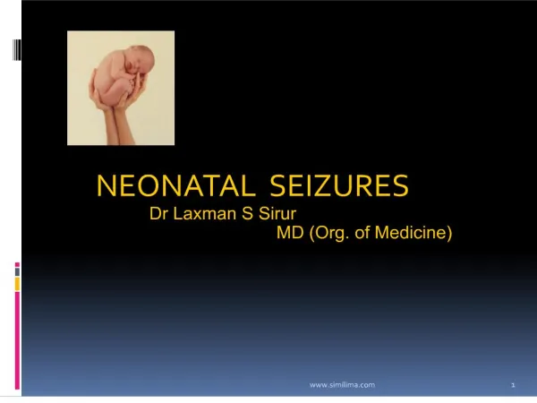
Neonatal Seizures Priscilla Joe, MD Children’s Hospital & Research Center at Oakland
Pathophysiology • Abnormal synchronous depolarization from large group of neurons • Excessive excitatory amino acid release (glutamate) • Lack of inhibitory systems (GABA) • Depolarization results from Na influx into cells; repolarization from outflux of K+ • Disruption of Na/K ATP pump
Basic Mechanisms of Seizures • Abnormal energy production (hypoxemia, hypoglycemia) • Alteration in neuronal membrane (hypocalcemia, hypomagnesemia) • Relative excess of excitatory versus inhibitory neurotransmitters (GABA)
Biochemical Changes with Seizures • ↓ ATP • ↓ phosphocreatine • Pyruvate converted to lactate • ↓ brain glucose • Increased production of pyruvate from ADP
Incidence • Higher in neonates than any other age group • Most frequent in the first 10 days of life
Do Prolonged Seizures Harm the Developing Brain? • Animal studies: • Persistent neonatal seizures in rats induce neuronal death and changes in hippocampus • Chronic seizures in adults associated with memory impairment and poor psychosocial outcome • Permanent reduction in seizure threshold associated with significant deficits in learning and memory
Causes of Neonatal Seizures • HIE (32%) • Intracranial hemorrhage (17%) • CNS infection (14%) • Infarction (7%) • Metabolic disorders (6%) • Inborn errors (3%) • Unknown (10%) • Drug withdrawal (1%)
Adverse Effects of Seizures • Cell division and migration • Formation and expression of receptors • Synaptogenesis and apotosis • Long term effects: seizure threshold, learning, and cognition
Subtle Seizures • More common in premature infants • Most frequently observed type of seizure • Clinical manifestations: Bicycling movements, lip smacking, apnea, and eye movements or staring, unresponsiveness • Typically have no electrographic correlate, are likely primarily subcortical
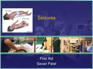
Clonic Seizures • Focal or multifocal, rhythmic movements with slow return movement • May be associated with generalized or focal brain abnormality • Most commonly associated with electrographic seizures
Tonic Seizures • Sustained flexion or extension of one extremity or the whole body • Extensive neocortical damage with uninhibited subcortically generated movements • May or may not have electrographic correlate
Myoclonic Seizures • Rapid, isolated jerks which lacks the slow return phase of clonic movements • Typically not associated with electrographic correlate • Myoclonic movements may be normal in preterm or term infants
Nonepileptic movements • Benign sleep myoclonus • Tremulousness or jitteriness • Stimulus evoked myoclonus from metabolic encephalopathies, CNS malformation
Benign Sleep Myoclonus • Onset 1st week of life • Synchronous jerks of upper and lower extremities during sleep • No EEG correlate • Provoked by benzodiazepines • Ceases upon arousal • Resolves by 2 months • Good prognosis
Jitteriness vs. Seizures • No ocular phenomena • Stimulus sensitive • Tremor • Movements cease with passive flexion
Hypoxic Ischemic Encephalopathy • Seizures begin within 24-72 hours after birth • Accounts for 50-60% of all neonatal seizures • Most asphyxia occurs before or during birth • Arterial cord pH < 7.0, base deficit < -10 • 60% develop seizures within 1st 12 hours • Recent stress: hypotonia and unresponsiveness • Longer standing dysfunction: hypertonia with cortical thumbing, joint contractures or conversely hypotonia with encephalopathy
Meningitis/ Encephalitis • Accounts for 5-10% of all neonatal seizures • TORCH, enterovirus, parvovirus Usually present by day 3 of life, except for HSV which may present in 2nd week of life • GBS, listeria, E coli, strep pneumoniae Presents at end of 1st week to 3 months of age
Intracranial Hemorrhage • Accounts for 10% of all seizures • Grade IV IVH/PVH • Subarachnoid/subdural hemorrhage • Cerebral infarction (ischemia, dehydration, infection, polycythemia)
Cerebral Infarction • Most frequently involves middle cerebral artery • Focal, clonic seizures common • At risk for spastic hemiparesis • Venous sinus thrombosis may cause hemorrhage stroke • ECMO
Etiologies: CNS malformations • Lissencephaly, pachygyria, linear sebaceous nevus syndrome, polymicrogyria • Present with seizures at a later age
Etiologies: Metabolic • Hypoglycemia, hypocalcemia, hypomagnesemia, hyper/hyponatremia • Inborn errors of metabolism (>72hrs of age): Aminoacidopathies, urea cycle disorders, biotinidase deficiency, mitochondrial disorders, beta oxidation disorders, glucose transporter deficiency, peroxisomal disorders
Epileptic syndromes-benign • Benign familial neonatal seizures • Autosomal dominant • Inter-ictal exam is normal • Long term outcome is good • Unusual tonic-clonic pattern • Benign idiopathic neonatal seizures • Term, normal birth • Normal inter-ictal state, EEG • Clonic, occur day 5, may be Zn deficiency
Epileptic syndromes-malignant • Neonatal Myoclonic encephalopathy • Fragmentary partial seizures, massive myoclonus • Metabolic disorders, abnormal EEG • Poor prognosis • Ohtahara syndrome • 10d -3 mo • Numerous brief Tonic seizures • Dysgenesis is cause, prognosis very poor
Metabolic Evaluation • Blood: glucose, lytes, BUN, creatinine, lactate, pyruvate, ammonia, biotinidase, quantitative amino acids, very long chain fatty acids • Urine: quantitative amino acids • CSF: cell count, glucose, protein, pyruvate, lactate, quantitative amino acids, HSV PCR
EEG • Scalp recordings measure discharges that spread to the surface • Discharges from frontal or temporal regions may not spread to the surface • More common in the newborn
Clinical Seizures Without EEG Correlate • May represent uninhibited brainstem reflexes • Discharges from deep cerebral structures and brainstem may not reach the cortical surface
Treatment • More difficult to suppress than in older children • Treatment is worthwhile because seizures: • May cause hemodynamic or respiratory compromise • Disrupt cerebral autoregulation • May result in cerebral energy failure and further injury
Treatment • Stabilize vital signs and treat underlying hypotension • Correct transient metabolic disturbances • Phenobarbital is first line agent • Lorazepam • Phenytoin
Prognosis based on etiology • Hypoxia-ischemia • Meningitis • Hypoglycemia • Early Hypocalcemia • Subarachnoid hemorrhage • Late Hypocalcemia 50% normal outcome Almost all are normal
Prognosis based on etiology • Cerebral dysgenesis has grave prognosis, almost none are normal • Prematurity and seizures associated with high risk of death or very poor outcome
Prognosis based on type • Subtle Depends on cause, other seizure types • Clonic Better prognosis • Generalized Tonic Poor • Myoclonic Poor
Prognosis by EEG • Severe inter-ictal EEG background associated with adverse outcome • Normal EEG background at presentation associated with good outcome • Ictal features less reliable • Better outcome when clinical and EEG seizures correlate • Increased number and frequency may relate to worse outcome
Conclusions • Neonatal seizures are often subtle • Close examination and characterization important for prognosis and evaluation • Treatment usually successful in stopping seizures, but risk of neuro-developmental abnormalities remains high • Prevention of causes remains a priority