Download
1 / 32
370 likes | 968 Vues
YEASTS Candida albicans Cryptococcus neoformans Prof. Khaled H. Abu-Elteen. Yeast. Yeasts are single-celled budding organisms . They do not produce mycelia . The colonies are usually visible on the plates in 24-48 h. Their soft, moist colonies

E N D
YEASTS Candida albicans Cryptococcus neoformans Prof. Khaled H. Abu-Elteen
Yeast • Yeasts are single-celled budding organisms. They do not produce mycelia. The • colonies are usually visible on the plates in 24-48 h. Their soft, moist colonies • resemble bacterial cultures rather than molds. There are many species of yeasts which • can be pathogenic for humans. We will discuss only the two most significant species: Candida albicans and Cryptococcus neoformans.
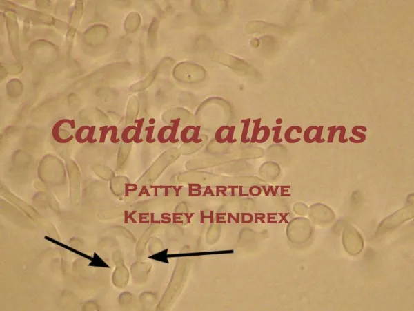
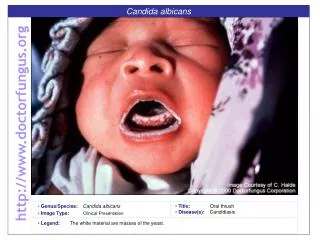
CANDIDIASIS (Candida albicans) There are many species of the genus Candida that cause disease. The infections caused by all species of Candida are called candidiasis. Candida albicans is an endogenous organism. It can be found in 40-80% of normal human beings. It is present in the mouth, gut, and vagina. It may be present as a commensal or a pathogenic organism. Infections withCandida usually occur when a patient has some alteration in cellularimmunity, normal flora or normal physiology.
Patients with decreased cellular immunity have decreased resistance to fungal infections. Prolonged antibiotic or steroid therapy destroys the balance of normal flora in the intestine allowing the endogenousCandidato overcome the host. Invasive procedures, such as cardiac surgery and indwelling catheters, produce alterations in host physiology and some of these patients developCandida infections
Although it most frequently infects the skin and mucosae, Candida can cause pneumonia, septicemia or endocarditis in the immuno-compromised patient. The establishment of infection withCandida species appears to be a property of the host - not the organism. The more debilitated the host,the more invasive the disease.
The clinical material to be sent to the lab depends on the presentation of the disease: blood cultures, vaginal discharge, urine, feces, nail clippings or material from cutaneous or mucocutaneous lesions. Candida is a polymorphic yeast, i.e., yeast cells, hyphae and pseudohyphae are produced. It has been shown that Candida needs a transcription repressor to maintain the yeast form.
This ability to assume various forms may be related to the pathogenicity of this organism. The yeast form is 10-12 microns in diameter, gram positive, and it grows overnight on most bacterial and fungal media. It also produces germ tubes, and pseudohyphae may be formed from budding yeast cells that remain attached to each other. Spores may be formed on the pseudomycelium. These are called chlamydospores and they can be used to identify different species of Candida.
Some mycologists think that the pseudomycelial form represents a more invasive form of the organism. The species are identified by biochemical reactions. The organism occurs world-wide. The drugs of choice for systemic infection are itraconazole and fluconazole. If anartificial heart valve orin-dwelling catheter becomes infected, itmust be replaced. Drug therapy alone will not suppress the organism if the foreign body remains in the host.
GERM TUBES GERM TUBES
Yeasts are single-celled budding organisms. They do not produce mycelia. The colonies are usually visible on the plates in 24-48 h. Their soft, moist colonies resemble bacterial cultures rather than molds. There are many species of yeasts which can be pathogenic for humans. We will discuss only the two most significant species: Candida albicans and Cryptococcus neoformans Parameters affecting adhesion of Candida species to epithelial cells, plastic surfaces and denture-base resins
Putative Virulence Factors of C. albicans • Adherence • Dimorphism • Germ tubes • Rapid switching of expressed phenotype • Surface hydrophobicity • Interference with phagocytosis, immune defences and complement • Extracellular hydrolases (Proteinases, lipases) • Synergism with certain bacteria • Killer toxins • Acidic metabolites • Growth rate and undemanding nutrient requirement
CRYPTOCOCCOSIS (Cryptococcus neoformans) Cryptococcosis manifests itself most commonly as meningitis but in recent years many cases of pulmonary disease have been recognized. C. neoformans is a very distinctive yeast. The cells which are spherical and 3-7 microns in diameter, produce buds which characteristically are narrow-based and the organism is surrounded by a polysaccharide capsule.
There is evidence that the capsule may suppress T-cell function and can be considered a virulence factor. C. neoformans also produces an enzyme called phenoloxidase which appears to be another virulence factor. The ecological niche ofC. neoformans ispigeon and chicken droppings. However,although this organism can be easily recovered from pigeon droppings, a direct epidemiological link has yet to be established between exposure to pigeon droppings and a specific human infection.
The source of human infection is not clear. The portal of entry is therespiratory system. Evidence is developing which indicates that the initial exposure may be many years prior to the manifestation of disease. The organism can be sequestered for this time. Infection may be subacute or chronic. The highly fatalmeningoencephalitiscaused byC. neoformans has a prolonged evolution of several months.
The patients symptoms may begin with vision problems and headache, which then progress to delirium, nuchal rigidity leading to coma and death unless the physician is thinking about cryptococcus and does a spinal tap for diagnosis and institutes aggressive therapy. The CSF is examined for its characteristic chemistry ) elevated protein and decreased glucose(, cells (usuallymonocytes), and evidence of the organism.
The latter is measured by the visual demonstration of the organism (India Ink preparation) or by a serologic assay for the antigen ofC. neoformans. The India Ink test, whichdemonstrates the capsule of this yeast, is supplemented by thelatex agglutinationtest for antigen which is more sensitive and more specific. The Latex Agglutination test measures antigen, NOT antibody.A decreasing titer indicates a good prognosis, while an increasing titer has a poor prognosis.
When you consider Cryptococcosis, think of Capsules and CNS disease. In addition to causing meningitis, C. neoformans may also infect lungs and skin. The disease in thelungs and skin is characterized by the formation of agranulomatous reaction with giant cells. As with other fungal diseases, there has been an increase in the recognition of pulmonary infection. The geographical distribution of this organism is world-wide.
The clinical material sent to the lab is CSF, biopsy material, and urine (for some unexplained reason the organism can be isolated from the urine in both the CNS and systemic infections). This organism will grow overnight on bacterial or fungal media at 37º C, but growth is a little slower at room temperature. In culture the organism grows as creamy, white, mucoid (because of the capsule) colonies. Growth in culture is usually visible in 24 to 48 h. As the culture ages, it turns brown due to a melanin produced by the phenoloxidase.
The organism is a round, single cell, yeast surrounded by a capsule. Identification is based on physiological reactions. • Pathologists use a muci-carmine stain, which stains the capsule, to identify the organism in tissue sections. There is usually little or no inflammatory response. The Direct Fluorescent Antibody test identifies the organism in culture or tissue section specifically, by causing the yeast cell wall to stain green.
To test the patient's serum there are 3 serologic tests: 1-The Indirect Fluorescent Antibody test, 2- the Tube Agglutination test for antibody, 3- the Latex Agglutination test for antigen. The latex agglutination test can be used as a prognostic test. As the patient improves, the serum antigen titer will also decrease. The drugs of choice to treat cryptococcus infection are amphotericin B and 5-Fluorocytosine (5-FC). 5-FC is an oral drug. If it is given as the only treatment, there are relapses so most physicians use both drugs simultaneously. Actually, these two drugs are synergistic, and thus, their association is advantageous.