Introduction :
Suspicious topography in family members of a patient with Pellucid Marginal Degeneration: a case for inheritance? Hajirah Saeed, M.D., Christine Garcia, B.S., Charles Bouchard, M.D. Loyola University Medical Center, Department of Ophthalmology, Maywood, Illinois .

Introduction :
E N D
Presentation Transcript
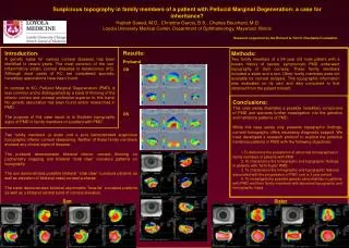
Suspicious topography in family members of a patient with Pellucid Marginal Degeneration: a case for inheritance? Hajirah Saeed, M.D., Christine Garcia, B.S., Charles Bouchard, M.D. Loyola University Medical Center, Department of Ophthalmology, Maywood, Illinois Research supported by the Richard A. Perritt Charitable Foundation Results: Proband OD OS Introduction: A genetic basis for various corneal diseases has been identified in recent years. The most common of the non-inflammatory ectatic corneal diseases is keratoconus (KC). Although most cases of KC are considered sporadic, hereditary associations have been found. In contrast to KC, Pellucid Marginal Degeneration (PMD) is less common and is distinguished by a band of thinning of the inferior cornea and corneal protrusion superior to this band. No genetic association has been found and/or researched in PMD. The purpose of this case report is to illustrate topographic signs of PMD in family members of a patient with PMD. Methods: Two family members of a 54 year old male patient with a known history of severe, symptomatic PMD underwent topography of their corneas. These family members included a sister and a son. Other family members were not available for corneal analysis. This topographic information was evaluated on its own and also compared to that obtained from the patient himself. Conclusions: This case series illustrates a possible hereditary component of PMD and warrants further investigation into the genetics and heritance patterns of PMD. While this case series only presents topographic findings, corneal tomography offers necessary diagnostic support. We have developed a research protocol to explore the potential heritance patterns in PMD with the following objectives: 1.To determine the prevalence of abnormal tomographies in family members of patients with PMD 2. To characterize the tomographic and topographic findings in patients with “form fruste” PMD 3. To characterize the tomographic and topographic features associated with the progression of PMD over a 3 year period 4. To investigate the possible genetic abnormalities in patients with PMD and their family members with abnormal topographic and tomographic maps Two family members (a sister and a son) demonstrated suspicious topographic inferior corneal steepening. Neither of these family members showed any clinical signs of disease. The proband demonstrates bilateral inferior corneal thinning on pachymetry mapping and bilateral “crab claw” curvature patterns on topography. The son demonstrates possible bilateral “crab claw” curvature patterns as well as elevation of bilateral nasal corneal surfaces. The sister demonstrates bilateral asymmetric “bow-tie” curvature patterns as well as a bilateral central band of corneal elevation. Son Sister