Sarcoidosis
Sarcoidosis. T. Lianne Beck, MD Assistant Professor Emory Family & Preventive Medicine. Objectives. Epidemiology Pathogenesis Clinical presentation Organ systems involved Diagnostic evaluation Current evidence on treatment. Sarcoidosis.

Sarcoidosis
E N D
Presentation Transcript
Sarcoidosis T. Lianne Beck, MD Assistant Professor Emory Family & Preventive Medicine
Objectives • Epidemiology • Pathogenesis • Clinical presentation • Organ systems involved • Diagnostic evaluation • Current evidence on treatment
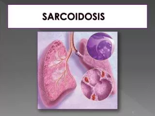
Sarcoidosis • Multisystem disorder of unknown etiology that most commonly affects the lungs, but can also affect other organs. • Beethoven is thought to have been the first person described with this condition.
Epidemiology • 3rd or 4th decade of life. • More predominant in women with an incidence of 6.3 vs 5.9 cases per 100,000 person-years. • Lifetime risk for US whites is 0.85 percent compared with 2.4 percent in US blacks. • More prevalent in Swedes, Danes, and US blacks.
Epidemiology • Annual incidence in the U.S. is 10/100,000 among whites and 36/100,000 among African Americans. • Most commonly seen in the mid-Atlantic and Southern Atlantic states but rare in the Southwest. • Affects siblings of first- or second- degree relatives in 15% of patients with sarcoidosis. • Familial cases described in 17% of African Americans, but only 6% of whites.
Etiology and Pathogenesis • Cause is unknown, although both genetic and environmental factors suspected. • Theory that disease develops in genetically predetermined hosts who are exposed to certain environmental agents that trigger an exaggerated inflammatory immune response leading to granuloma formation.
Etiology and Pathogenesis • Hallmark is noncaseating granulomas, composed of a central core of epithelioid histocytes and multinucleated giant cells. • Activated T cells and macrophages accumulate at site of inflammation. • Release chemoattractants and GF’s lead to cellular proliferation and granuloma formation. • Progressive granulomatous inflammation leads to injury, dysfunction, and destruction of the affected organs.
Pathogenesis T cells, Macrophages Chemoattractants Growth Factors Cellular proliferation Granuloma Fibrosis
Clinical Presentation • 30-50% of patients are asymptomatic and are diagnosed on routine CXR. • One third have non-specific symptoms of fever, fatigue, weight loss and malaise. • A clinical variant of sarcoidosis, Lofgren’s syndrome, includes constellation of erythemanodosum, polyarthritis, and BHL. Remission occurs in 80%.
Clinical Presentation • Onset of sarcoidosis in white patients is usually asymptomatic. • African Americans tend to present with an earlier onset and a more aggressive and severe clinical course. • Chronic pulmonary sarcoidosis and the disfiguring cutaneous lesions of lupus pernio are also more common in African Americans.
Clinical Presentation • Spontaneous remission in two-thirds of patients within 2 years of presentation • 10%-30% experience chronic disease causing progressive organ damage • Leads to death in 4% of patients, usually those with pulmonary, cardiac, or CNS involvement
Systems affected by Sarcoidosis Signs and symptoms
Systems affected by Sarcoidosis Signs and symptoms
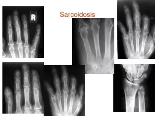
Clinical Presentation • A progressive course is more likely in: • Age of onset > 40 yrs • Black race • Cardiac or renal involvement • Lupus pernio • Chronic uveitis • Hypercalcemia • Nasal mucosal involvement • Cystic bone lesions • Neurosarcoidosis • Pulmonary fibrosis
Clinical Presentation • Most patients have the pulmonary manifestations, most commonly presenting with incidental findings on CXR. • Interstitial disease • Symptoms include dry cough, dyspnea, and chest discomfort • Unpredictable course
Approach to Suspected Sarcoid • History (occupational and environmental) • PE (lungs, skin, eyes, liver, and heart) • CXR, PFT’s and EKG • CBC, CMP, ACE level • PPD • Biopsy for histological confirmation of noncaseatinggranulomas and culture and/or special staining to R/O fungal or TB • Ophthalmologic evaluation
Approach to Suspected Sarcoid • Follow-up • Monitor for resolution or progression of disease and for additional organ involvement. • Refer if there is evidence of disease progression or additional organ involvement. • Coordinate care.
Approach to Suspected Sarcoid • An aggressive work up may be unnecessary in asymptomatic patients with symmetric BHL, unremarkable exam, no history of malignancy, and normal results on routine bloodwork. • The course of disease usually becomes evident within 2 years of presentation. Absence of remission within this period predicts a chronic, persistent, or stable course.
Differential Diagnosis of BHL • Granulomatous infections • TB • Histoplasmosis • Coccidiomycosis • Autoimmune disorders • Malignancy (Lymphoma)
Differential Diagnosis of Noncaseating Granulomas • TB • Fungal infections • Lymphoma • Epithelioid tumors of the breast • Lung cancer
Treatment • Observation • Initiating corticosteroid therapy when appropriate • Monitoring response to therapy • Discontinuing corticosteroids when clinically or physiologically indicated.
Treatment • Topical therapy for cutaneous or ophthalmic disease. • Systemic corticosteroids for patients with unresponsive ophthalmic manifestations, cardiac, neurologic and progressive pulmonary involvement. • Systemic therapy for patients with hypercalcemia.
Treatment • Prednisone, 20 to 40 mg/d in divided doses or alternate-day dosing is used for organ involvement that is not life threatening. • Higher dosage is used off-label for potentially life threatening disease. • High-dose inhaled corticosteroids may be useful in patients with symptomatic pulmonary disease.
Treatment • Clinical improvement should be assessed after 3 months of corticosteroids. • If no improvement is found, further treatment is unlikely to be beneficial. • Long term adverse affects of therapy include weight gain, mood swings, cataracts, GERD, osteoporosis