Veterinary Radiography Techniques for Small Animal Forelimb
300 likes | 346 Vues
Learn methods to radiograph the scapula and forelimb of small animals with detailed instructions for positioning the patient and image capture. Techniques include dorsal and cranial views for optimal diagnostics.

Veterinary Radiography Techniques for Small Animal Forelimb
E N D
Presentation Transcript
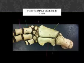
Small Animal Forelimb Ch. 13
Scapula • Two methods of radiographing the Scapula exists. • 1. With the scapula placed dorsal to the vertebral column • 2. With the scapula superimposed over the lung fields
Scapula Dorsal to the Vertebral Column • Push the leg dorsally so the scapula is positioned dorsal to the vertebral column. • Patient is in lateral recumbency with shoulder of interest closest to the cassette. • Push limb dorsally with elbow extended. • Sternum may be slightly rotated if needed. • Beam should be centered over middle of scapula.
Scapula Superimposed over cranial Thorax • When patient is in pain or when manipulation may cause further injury. • Body of scapula is over translucent lung fields, allowing visualization of the majority of the bone. • Area of interest closest to cassette, upper limb extended cranially and out of field.
Caudocranial View of Scapula • Patient is in dorsal recumbency with both forelegs extended cranially.
Shoulder- Lateral view • Area of interest closest to the cassette. • Leg extended cranially and ventrally to sternum. • Opposite leg pulled back and out of the way.
Shoulder- Caudocranial view • Similar to that of scapula. • Leg should be extended until forelimb is almost parallel to cassette. • Sometimes can get comparison view of both shoulders on this view.
Humerus- Lateral View • Lateral recumbency with area of interest closest to cassette. • Leg is extended forward and opposite leg drawn back. • Head and neck should be up and out of field of view.
Humerus- Caudocranial view • Patient in Dorsal recumbency with forelimbs extended cranially. • Leg of interest parallel to cassette. • Humerus centered to the cassette. • See example in textbook.
Humerus-Craniocaudal View • Patient in Dorsal Recumbency • Area on interest pulled caudally until humerus is parallel with cassette. • Field of view should include shoulder, humerus, and elbow.
Elbow- Craniocaudal View • Patient in sternal recumbency with limb extended cranially. • Head should be elevated and positioned away from the affected side. • Try to avoid rotation.
Elbow- Lateral View • Patient in Lateral Recumbency with area of interest closest to the cassette. • Head and neck should be extended slightly dorsally. • Unaffected limb should be pulled back.
Elbow- Flexed Lateral View • Same positioning as routine lateral view. • Elbow is simply flexed.
Radius and Ulna- Lateral View • Patient in Lateral Recumbency. • Limb centered on cassette. • Should get joint above and joint below.
Radius and Ulna- Craniocaudal View • Patient is in sternal recumbency. • Limb is extended cranially with radius and ulna centered on the cassette.
Carpus- Lateral View • Affected limb on center of cassette. • Patient in Lateral Recumbency. • Opposite limb is pulled out of field of view.
Carpus- Dorsopalmar View • Patient is in sternal Recumbency with affected limb cranially. • Carpus is placed flat on cassette.
Metacarpus-Phalanges Dorsopalmar View • Patient is in sternal recumbency with limb extended. • Paw is flat on cassette.
Metacarpus-Phalanges Lateral View • Patient is in Lateral Recumbency with affected side down. • Limb of interest on cassette. • Results may be that phalanges are superimposed on one another.