Download
1 / 25
250 likes | 269 Vues
This case report discusses a 20-year-old male presenting with back and abdominal pain, showcasing radiological findings of retroperitoneal masses. The differential diagnosis includes lymphoma, metastases, sarcoma, and hemangiopericytoma.

E N D
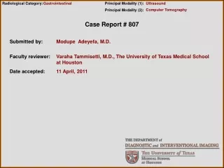
Radiological Category: Gastrointestinal Principal Modality (1): Principal Modality (2): Ultrasound Computer Tomography Case Report # 807 Submitted by: Modupe Adeyefa, M.D. Faculty reviewer: Varaha Tammisetti, M.D., The University of Texas Medical School at Houston Date accepted: 11 April, 2011
Case History 20 year old male with a three month history of back pain relieved by Aleve but the pain has progressively worsened. He went to his primary care physician where a right lower quadrant mass was palpated. An abdominal ultrasound was performed which showed two large retroperitoneal masses. He presented about a week later to Memorial Herman Hospital because of worsening abdominal pain and syncope.
Ultrasound: Two large heterogeneous echogenic retroperitoneal masses.CT: Large intraperitoneal hemoperitoneum and large “sentinel clot” adjacent to the necrotic, hypervascular nodal masses in lower abdomen. There are 3 large, centrally necrotic, peripherally enhancing masses in the retroperitoneum, the largest in the pelvis and measures 9.58x8.8 cm. Smaller similar masses are noted further superiorly . Findings and Differentials Findings: Differentials: • Lymphoma • Retroperitoneal Metastases with tumor associated hemorrhage/ hemoperitoneum • Sarcoma • Retroperitoneal Hemangiopericytoma
Lymphoma: Accounts for the 10%-15% of all childhood cancer which arises from constituent cells of the immune system or from precursors. Nodal and splenic involvement are more common in Hodgkin disease whereas extranodal involvement is more common in Non Hodgkin lymphoma. Both can involve the retroperitoneum. It appears as a mantle of soft tissue adenopathy surrounding the aorta, inferior vena cava, involving the para-aortic, aortocaval and retrocaval nodal groups. CT findings are enlarged nodes greater that 1.5 cm in the short axis involving bilateral retroperitoneal nodal chains. Encasement of the root of the mesentery and the superior mesenteric artery by a lymphomatous mass originating from multiple enlarged and confluent lymph nodes may produce the so called “sandwich sign.” They may displace aorta from the spine. The attenuation of nodes on CT is similar to muscle. Hemoperitoneum from pathologic splenic rupture is rarely associated with lymphoma in which case the sentinel clot would be seen in the perisplenic region. In this patient the sentinel clot is seen surrounding the lymph nodes and the lymph nodes are necrotic with hypervascularity, both of which are not common features associated with lymphoma. Discussion
Retroperitoneal Metastasis with tumor associated hemorrhage: Spontaneous hemoperitoneum rarely occurs in the absence of trauma, a surgical or interventional procedure, or anticoagulation therapy. In such cases, the possibility of the rupture of an un-identified neoplasm must be excluded. Although the occurrence is uncommon, any primary or metastatic tumor can rupture and bleed into the peritoneal cavity. The most common causes in this setting would be ruptured hepatocellular carcinoma or hepatocellular adenoma. However, no hepatic lesions are identified and sentinel clot is seen away from the liver. Pathologic splenic rupture may occur as a complication of a viral infection, including infection by cytomegalovirus, malaria, or Epstein-Barr virus; a congenital disease; a metabolic abnormality such as Gaucher disease or amyloidosis; and, rarely, a neoplastic process such as hemangiomatosis, angiosarcoma, leukemia, or lymphoma. However, the sentinel clot is not in the perisplenic region and there is no splenomegaly. The spontaneous rupture of a metastatic lesion in a solid organ or lymph node is rare but usually results in massive hemoperitoneum. Lung carcinoma, renal cell carcinoma, and melanoma are the metastatic lesions that can cause hemoperitoneum. Discussion
Sarcoma: Malignant primary retroperitoneal tumor arising from various elements of primitive mesenchyme, urogenital ridge or embryonic remnants. The best diagnostic clue on imaging is a large heterogenous mass of fat and soft tissue attenuation displacing the retroperitoneal structures or viscera. The location is usually in the peri/paranephric region. They are classified into four types, liposarcoma, leiomyosarcoma, fibrosarcoma or rhabdomyosarcoma. Liposarcoma is composed of adipose tissue with attenuation values greater than 20 hounsfied unit. They are poorly marginated encapsulated masses with/without calcification. On CT they have hetero/homogenous enhancement with a lack of prominent vessels. Leiomyosarcoma is the second most common and are composed mostly of smooth muscle. They are hypervascular and contain feeding vessels. They may appear as large solid masses with hypoechoic cystic and necrotic remnants. Fibrosarcoma and malignant histiocytoma are the most common soft tissue sarcomas in adults. Rhabdomysarcoma are striated muscle tumors . They are isodence to muscle with ill defined margins on non enhanced CT and are heterogenously enhancing on contrast enhanced CT. Discussion
Retroperitoneal Hemangiopericytoma are hypervascular neoplasm arising form pericytes. In a study by Goldman et al, these tumors were typically large and less frequently in pelvic retroperitoneal space than in abdominal retroperitoneal. The most distinctive radiologic finding is hypervascularity, Other findings includes well defined margins and necrosis with nondistinctive amorphous calcifications. Discussion
Additional Images Multiple pulmonary nodules with halo of ground glass haziness “halo sign”
The halo sign, refers to a zone of ground glass attenuation surrounding a pulmonary nodule or mass on CT images and they are associated with hemorrhagic nodules, examples include: Angio-invasive aspergillosis: is a form of tissue invasion either angioinvasive or airway invasive typically in patients with neutropenia or impaired neutrophil function. Imaging findings include single or multiple nodules, a central hypodensity due to infarction also known as hypodense sign. The halo sign is also present which shows a large bull’s eye surrounded by smaller rim ground glass opacification. An air cresent sign is a late sign and follows recovery of neutrophils. Hemorrhagic/ hypervascular metastases such as from choriocarcinoma, renal cell carcinoma, angiosarcoma or melanoma. The nodules may be sharply defined. These findings mirrors chest x-ray findings with metastases predominate in the outer 1/3 of the mid and lower lung zones. Hematogenous nodules may have visible feeding artery or the halo sign. Discussion
Wegener granulomatosis: A form of vasculitis that affects the lungs, kidneys and other organs. The best diagnostic clue are multiple cavitary lung nodules and large airwary narrowing. CT findings are similar to metastases as the nodules can have feeding vessels. Peripheral wedge shaped consolidation from infarct may be present. Some nodules have the CT halo sign from surrounding hemorrhage. The trachea and bronchi are concentrically thickened, either focal or long segments. Post transplant lymphoproliferative disorder (PTLD): it is a relatively uncommon complication of both solid organs and allogeneic bone marrow transplantation. In most cases, PTLD is associated with Epstein Barr virus infection of B cells, either as a consequence of a reactivation of the virus posttransplantation or from primary EBV infection acquired from the donor. CT findings include airspace consolidation and nodular opacities with hazy margins. Pulmonary Kaposi sarcoma: Acquired immune deficiency syndrome related multicentric neoplasm with propensity to involve skin, lymph nodes, GI tract and lungs. Thoracic manifestations include bronchovascular bundle thickening progressing to coalescent, flame shaped perihilar consolidation, poorly defined nodules, reticular and nodular opacities with basilar predominance. There is also marked enhancement following intravenous contrast. Discussion
Additional Images Heterogeneous right testicular mass with cystic focus, few tiny calcifications, vascularity and irregular margins.
Based on the findings from the retroperitoneal masses, positive halo sign on CT and a testicular mass, the diagnosis is more likely an hemorrhagic or hypervascular metastases from a non-seminomatous germ cell tumor such as from choriocarcinoma or mixed germ cell tumor. Differential
METASTATIC MIXED OR NONSEMINOMATOUS GERM CELL TUMOR (NSGCT) SUCH AS CHORIOCARCINOMA Diagnosis
Pathology report from biopsied retroperitoneal masses. - Immunohistochemical stains for Pancytokeratin, Vimentin, Beta-HCG, Alpha Feto-Protein, CD30, Placental Alkaline Phosphatase, and Desmin were applied to the cell block. The positive stains are Beta HCG with the rest being negative. This supports the diagnosis of a choriocarcinoma. - Morphologically and immunohistochemically, the tumor was a choriocarcinoma. However the retroperitoneal mass is large, therefore a mixed germ cell tumor cannot be excluded. Discussion
Testicular Cancer is relatively uncommon in the United States with approximately 5500 cases per year. These tumors arises in males of nearly any age and may be of germ cell or non germ cell origin. There are three main types, germ cell tumors, non germ cell tumor and extragonadal tumors. Discussion
Choriocarinoma is a rare germ cell tumor and in its pure form , it is seen in less than 1% of patients, but it occurs in mixed cell tumors in 8% of cases. It typically affects younger men and unlike other cancers, choriocarcinoma metastasizes hematogenously, with the testicular primary tumor often small or even “burnt out.” It metastasizes early via hematogenous routes to the lung, liver, and brain, among others. This tumor represents the most common malignancy in men between the ages of 15-35 years of age. The tumor responds poorly to radiation and chemotherapy and carries high mortality rate. Patients with mixed germ cell tumors with choriocarinoma fair better than those with pure choriocarcinoma tumors, but a high human chorionic gonadotropin (>50,000 IU/L) portends a poor prognosis with at 5 year survical rate of 48%. Surgery is usually limited to radical orchiectomy. Diagnosis
After four rounds of chemotherapy, the patient had a right radical orchiectomy which showed a mixed germ cell tumor consisting of mature teratoma (approximately 95%) and seminoma (less than 5%). No residual choriocarcinoma was seen which indicates excellent response to therapy. Discussion
Goldman SM, Davidson AJ, Neal j. Retroperioteneal and pelvic hemangiopericytomas: clinical, radiologic and pathologic correlation. Radiology. July 1988, 168: 13-17Meghan Lubner, MD, Christine Menias, MD, Creed Rucker, MD, Sanjeev Bhalla, MD, Christine M. Peterson, MD, Lisa Wang, MD and Brett Gratz, MD. Blood in the Belly: CT findings of Hemoperitoneum. Radiographics. January 2007. Volume 27, 109-125Paolo Toma, MD, Claudio Granata, MD, Andrea Rossi, MD, Alberto Garaventa, MD. Multimodality Imaging of Hodgkin Disease and Non-Hodgkin Lymphoma in Children. Radiographics. September-October 2007. Volume 27(5) 1335-1347Woodward et al. From the Archives of the AFIP, Tumors and Tumorlike Lesions of the Testis: Radiologic and Pathologic Correlation. January 2002. Radiographics, Vol 22, 189-216.Statdx.comwww.emedicine.com References


