The Brain and Behavior
The Brain and Behavior. McElhaney. Biopsychology= Bio explanation/cause for behavior. Or Physiological Psychology. Includes Electro/Chemical processes Brain Anatomy The Study of Biological Aspects of Behavior Hormonal aspects of behavior Hemisphere specialization.

The Brain and Behavior
E N D
Presentation Transcript
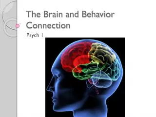
The Brain and Behavior McElhaney
Biopsychology= Bio explanation/cause for behavior. Or Physiological Psychology • Includes Electro/Chemical processes • Brain Anatomy • The Study of Biological Aspects of Behavior • Hormonal aspects of behavior • Hemisphere specialization
Phineas Gage- Frontal Lobe injury • Suffered brain injury that caused changes in his personality and behavior.
Brain Mapping • We know about the brain through experiments • Where portions of the brain are stopped, in order to identify locals of control.
What tools do Doctors have in determining Brain Function? • EEG • CAT Scan • MRI • PET Scan • Role of Glucose
Tools for Viewing Brain Structure and Activity • EEG • Electroencephalogram • measures electrical currents across the brain • Measure brain activity • And Location of different functions
Tools for Viewing Brain Structure and Activity • CT scan • Also called a CAT scan • Computerized axial tomography • X-ray of brain tissue • Shows brain structure • Cross section images
Snapshots of the Brain • PET scan • Positron Emissions Tomography • Patients drinks radioactive glucose* and image shows areas of brain activity or as it’s working. *Glucose is the primary fuel of the Brain as it is working. More glucose in an area means brain is working.
Tools for Viewing Brain Structure and Activity • MRI • Magnetic Resonance Imaging • Exposes brain to magnetic field • Shows brain structure
Neurons are the basic building blocks • AKA Nerve Cells – 100 Billion in brain • Carries and processes information • Connected via Chains of specailzed information • Activate Muscles & Glands • Non are identical
The Neuron and what it does • Dendrites= “Tree Roots” receives and messages from other neurons • Soma – cell body, sends Nerve Impulses • Axon- Fibrous carrier of impulses • Axon Terminals- end of Axon • Myelin Sheath- insulating body surrounding the axon
Nerve Impulse- “Firing” of a nerve • Ions= sodium (Na) Electrical Charged molecules are in each neuron • Electrical charge of <-70> Millivolts • Threshold = trigger point for sending a message • Nerve Impulse- will be fired when <-50> Millivolts are reached =Action Potential • Gates open and allow flow of ions- sodium goes in Potassium out
Psych Jeopardy Review • Choose group of 5 people • Divide Key Terms form reading (part 2 recommended) • Make questions/answers on flashcards (at least 10) • Play Jeopardy monday
Quiz Thursday: • Neuron and its components • Nerve Impulse and Action Potential • Synapse • Neurotransmitters
Synapses and Neurotransmitters • Info is transmitted chemically • Synapse = space between neurons • Between Axon terminals and Dendrites • Neurotransmitter=a chemical released by a neuron that stimulates nearby neurons and allows for nerve impulses to be passed throughout the body • Neurotransmitters are kept in vesicles, which fuse with the axon terminal’s membrane and travel into the synaptic cleft, ready to bind to receptors in the postsynaptic membrane
What a neurotransmitter looks like… Receptors are found at each end of the neuron- cell body and dendrite Neurotransmitters can slow or speed up firing of neuron There are over 100 transmitter chemicals
Facts and Such. • Everything you do or feel occurs due to communication between different neurons, which provide information throughout the nervous system. Within a single neuron, information travels through electrical signals, but when information is transmitted from one neuron to the next neuron, the transmission is considered ‘chemical’. • For two neurons to communicate neurotransmitters (messengers) are released into the synaptic cleft (an extremely tiny gap between neurons), where they then move to the next neuron and attach themselves to locations called receptorsites. • The result is an initiation of electrical current that moves through that neuron toward the next one. After the neurotransmitter does its thing, it is either destroyed by other chemicals in the synaptic cleft or is taken back into its original neuron. This action prevents the neurons from becoming ‘overstimulated’. • When neurons communicate, the effect can either help or hinder the next neuron. For example, when a person pays attention to one conversation and ignores others, the neurons in the brain are ‘listening’ to that information (helping) and ignoring the rest (hindering). Neurons come in different shapes and sizes, affecting many other neurons, and can have different numbers of synapses. Some neurons, called Purkinje cells, may have as many as 100,000 synapses.
Neurotransmitters Chemicals that influence the firing of nerve cells Can “excite”-make firing more likely Or “inhibit” make firing less likely
Effects of Drugs + Meds on Neurotransmitters • Alcohol- Increases GABA, Increases Dopamine • Prozac- Increases Serotonin levels • Same with Zoloft, Paxil, Celexa, Luvox • Opiates- Increases Dopamine, Mimics Endorphins • Morphine and heroin • Nicotine- Imitates Acetylcholine
Drugs and Neurotransmitters • Psychoactive medications: • Imitate • Duplicate • Or blocks Neurotransmitters
Curare = poison • Prevents Acetylcholine- up take • And causes paralysis
Endorphins • Endorphins are a group of small proteins naturally occurring in the brain around nerve endings that bind to opiate receptors • Natural opiates produced in the brain which function as the body’s own natural painkillers and Elevate mood. • Endorphins respond to morphine • Enkephalins= opiate like neural regulators relieve pain & stress similar to endorphins
Peripheral Nervous System • Controls the voluntary behavior • Carries information to and from the Central Nervous System • Peripheral nervous system is made up of: • Somatic System • Autonomic System • Sympathetic Branch • Parasympathetic Branch
Peripheral Nervous System • Somatic System- controls voluntary behavior • Sense organs and skeletal muscles
Sympathetic and Parasympathetic • Both systems work together: • Emotional responses and Involuntary behavior • Parasympathetic= quiets body, • returns to lower level of arousal, after emotional event • Vital functions, heart rate, breathing, digestion
Types of Neurons • Sensory Neurons – Afferent Neurons • Carry the message from the sense organs to the CNS • Inter neurons • Make up the CNS • Motor Neurons – Efferent Neurons • Carry the message from the CNS to the muscles or glands • Remember – SAME (sensory = afferent, motor = efferent)
Brain Stem • Medulla– where spinal cord meets the skull; controls heartbeat and breathing • Reticular formation– bundle of nerves running through the brainstem; controls arousal and attention; filters incoming stimuli and relays important information to the brain.
Cerebral Cortex Controls information processing; wrinkled to increase surface area Composed of 8 lobes (4 on each side)
Corpus Callosum bundle of nerves connecting the left and right hemispheres
Parietal Lobes Located on the top and rear of head Contains the sensory cortex (part of brain that registers and processes tactile information (phantom limb) Contains the angular gyrus (left hemisphere only) which is involved in converting written words into sound
Frontal Lobes Located in the forehead region Includes the motor cortex (part of brain that controls voluntary movement) • Includes Broca’s area (needed for forming words; located in left hemisphere only) • Association areas in this region – judgment, planning, processing new memories
Occipital Lobes • Located in the back of the head • Contains the visual cortex
Temporal Lobes Located on the sides of head, above ears Receives and processes auditory information Includes Wernicke’s area (left hemisphere only) - part of brain involved in understanding language
Cerebellum • Controls balance and coordination • In the rear of the head, behind the brainstem
Thalamus Thalamus • Pair of egg-shaped organs above the brainstem; receives information from the senses (EXCEPT FOR SMELL) and relays it to the rest of the brain.
Limbic System • Amygdala – two almond shaped structures; influence fear and aggression (monkeys and cats) • Hypothalamus – below the thalamus; regulates hunger, thirst, body temp, sex, fight-or-flight; triggers the pituitary (the “master gland”); reward center • Hippocampus – behind the amygdala; memory