Download
1 / 13
130 likes | 158 Vues
Surgical management of liver haemangioma. Dr Chris Yau Dept of Surgery PWH. 68 F USG: 3cm hyperechoic lesion right lobe Typical haemangioma Hepatitis marker –ve, AFP CEA normal FU CT scan: 7cm after 3 yr Trisegmentectomy: cholangiocarcinoma. 67 F USG: 4cm lesion left lobe

E N D
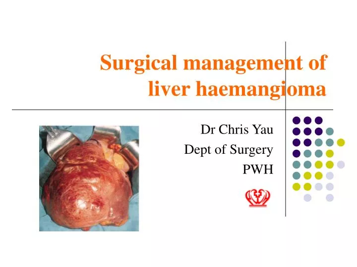
Surgical management of liver haemangioma Dr Chris Yau Dept of Surgery PWH
68 F USG: 3cm hyperechoic lesion right lobe Typical haemangioma Hepatitis marker –ve, AFP CEA normal FU CT scan: 7cm after 3 yr Trisegmentectomy: cholangiocarcinoma 67 F USG: 4cm lesion left lobe Typical haemangioma Hepatitis marker, AFP CEA normal FU USG 1 yr 8cm mass left lobe, multiple satellite lesions right lobe Palliative care Clinical scenario --- pitfall
Content • Pathology • Prevalence / Etiology • Clinical presentation • Diagnostic tools • Surveillance • Surgical management • Take home message
Liver Haemangioma --Pathology • First prescribed by Amboise Pare 1570 (Paris) • Benign tumour • Size up to 30cm • Giant Haemangioma ifsize > 10cm
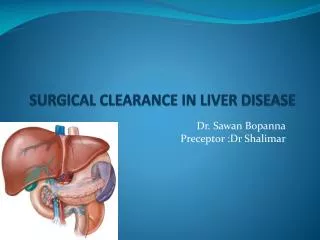
Liver Haemangioma --Pathology • Microscpically: • large endothelium lined vascular channels packed with red blood cell • Macroscopically: • red wine colored mass with lobulated appearance
Prevelance / Etiology • 0.7% to 7.3 % in autopsy finding * • Female predominance 1.5 – 6 : 1 ** • Present most often between 3rd and 4th decade • Positive correlation with female sex hormone *Belghiti, Management of haemangioma, Stuttgart: 1993: 78-85 ** Yan et al, US guided percutanceous RFA for hepatic haemangioma. World J Gastroenterol, 2003 Sept 2132-2134
Clinical Presentation • Asymptomatic • Epigastric pain • Spontaneous hemorrhage • Traumatic hemorrhage • Cardiac failure
Dignostic tools • USG: • Homogenous hyperechoic • CT scan: • Pre-contrast: Relative hypodense • Early phase : Perilesional increase in contrast • Delayed: isodense / hyperdense
Diagnostic tools • MRI scan: • T1 : hypointense • T2 : heavily hyperintense • Arteriography: • Golden standard • Cotton Wool appearance
Gandolfi (Italy) Nippon (Japan) Leifer (US) No of pt 123 23 383 Drop out NA Nil 170 Follow up period 12-60 months 12-114 months 12 yrs Change of lesion 1 Nil NA Malignancy Nil Nil 1 Complications Nil Nil NA Surveillance
Surgical Treatment • Treatment method: • Laparoscopic Vs Open • Resection Vs Enucleation • RFA • Indications of Surgery • Uncertain diagnosis • Symptomatic • Lesion with documented increase in size • +/- Giant haemangioma
Yoon et al Moreno et al Farges et al Ozden et No of pt 115 26 163 42 Surgery 52 15 8 42 Conservative tx 63 11 155 0 Malignancy 0 0 0 0 Morbidity 25% 13% NA 12% Mortality 0 0 NA 2.4% Surgery result
Take home message • Majority haemangioma can be treated conservatively • Existence of mimicker • Repeat scan / FNAC / Surgery if uncertain diagnosis