ABDOMINAL ASSESSMENT
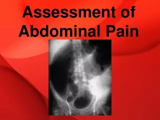
ABDOMINAL ASSESSMENT. Abdominal Assessment. Patient needs to be exposed from above the xiphoid process to the symphysis pubis. Also, make sure your patient does not have a full bladder. Place patient in a supine position: pillow under the head and knees. Helps to relax abdominal muscles.

ABDOMINAL ASSESSMENT
E N D
Presentation Transcript
Abdominal Assessment • Patient needs to be exposed from above the xiphoid process to the symphysis pubis. • Also, make sure your patient does not have a full bladder. • Place patient in a supine position: pillow under the head and knees. • Helps to relax abdominal muscles.
Abdominal Assessment • Have patient point out any areas of pain or tenderness. • Examine these last. • During exam continue to monitor your patient’s facial expression for pain and discomfort. • Use inspection, auscultation, percussion, and palpation to perform the exam.
Abdominal Assessment • Always auscultate before percussing or palpating. • These manipulations may alter your patient’s bowel motility and resulting bowel sounds.
Abdominal Assessment • Inspect the skin of the abdomen and flank’s for: • Scars • Dilated veins • Stretch marks • Rashes • Lesions • Pigmentation changes
Abdominal Assessment • Look for discoloration over the umbilicus: • Cullen’s Sign: discoloration over the umbilicus • Grey Turner’s Sign: discoloration over the flanks • These are both late signs suggesting intra-abdominal bleeding
Abdominal Assessment • Assess the size and shape of your patient’s abdomen to determine: • Scaphoid (concave) • Flat • Round • Distended • Ask the patient if it is its usual size and shape
Abdominal Assessment • Check for: • Bulges • Hernias • Distended Flanks • Ascites appears as bulges in the flanks and across the abdomen and indicates edema caused by CHF, or liver failure.
Abdominal Assessment • Look at your patient’s umbilicus • Note location and contour and observe for any signs of herniation or inflammation. • Check for: • Visible pulsation • Visible peristalsis (wavelike motion of organs moving their contents through the digestive tract). May indicate bowel obstruction. • Visible masses
Abdominal Assessment • Next auscultate for bowel sounds and other sounds such as bruits throughout the abdomen. • Gently place the diaphragm on your patient’s abdomen and proceed systematically, listening for bowel sounds in each quadrant. • Note location, frequency, and character • Normal bowel sounds consist of a variety of high-pitched gurgles and clicks that occur every 5-15 seconds.
Abdominal Assessment • More frequent sounds indicate increased bowel motility in conditions such as diarrhea or an early intestinal obstruction. • You may hear loud, prolonged, gurgling sounds known as borborygmi. • These indicate hyperperistalsis. • Decreased or absent sounds suggest a paralytic ileus or peritonitis
Abdominal Assessment • Bruits are swishing sounds that indicate turbulent blood flow. • Listen in areas over abdominal blood vessels such as the aorta and renal arteries • Presence indicates abdominal aortic aneurysm or renal artery stenosis
Abdominal Assessment • Percussing the abdomen produces different sounds based on the underlying tissues. • Sounds help you detect excessive gas and solid or fluid-filled masses • Also help you determine the size and position of solid organs such as the liver and spleen • Percuss the abdomen in the same sequence you used for auscultation
Abdominal Assessment • Note the distribution of tympany and dullness • Expect to hear tympany in most of the abdomen • Expect dullness over the solid abdominal organs such as the liver and spleen
Abdominal Assessment • Palpate the abdomen last to detect: • Tenderness • Muscular rigidity • Superficial organs and masses • Before you begin palpation, ask your patient if he has any pain or tenderness • Palpate that area last, using gentle pressure with a single finger
Abdominal Assessment • Ask him to cough and tell you if and where he experiences any pain • This is typical for peritoneal inflammation
Abdominal Assessment • Light palpation by moving your hand slowly and just lifting it off the skin. • Use same sequence as for auscultation and percussion • Watch for patient’s face for signs of discomfort
Abdominal Assessment • Identify any masses and note: • Size • Location • Contour • Tenderness • Pulsations • Mobility
Abdominal Assessment • Abdominal pain upon light palpation suggests peritoneal irritation or inflammation • If rigidity or guarding while palpating, determine whether it is voluntary (patient anticipates the pain) or involuntary (peritoneal inflammation)
Abdominal Assessment • Next palpate deeply to detect large masses or tenderness • Use one hand on top of another and push down slowly. • Assess for rebound tenderness by pushing slowly and then releasing your hand quickly off the tender area.
Abdominal Assessment • If you note a protruding abdomen with bulging flanks and dull percussion sounds in dependent areas, you might perform two tests for ascites.
Ascites/Test 1 • Assess for areas of tympany and dullness while your patient is supine • Lie him on one side • Percuss again, noting once more any areas of tympany and dullness • If your patient has ascites, the area of dullness will shift down to the dependent side and the area of tympany will shift up.
Ascites/Test 2 • Test for fluid wave, ask an assistant to press the edge of his hand firmly down the midline of your patient’s abdomen • With your fingertips, tap one flank and feel for the impulse’s transmission to the other flank through excess fluid • If you detect the impulse easily, suspect ascites