Download
1 / 49
500 likes | 880 Vues
LABORATORY SKILLS MIC-240. Dr. Kahkashan Perveen Bulding. 4, Floor 1, Room 415 E-mail: kperveen@ksu.edu.sa. Tests and Exams:. REPORTS 10 % QUIZ 20 % ASSIGNMENT 15 % CLASS PERFORMANCE 10 %

E N D
LABORATORY SKILLSMIC-240 Dr. Kahkashan Perveen Bulding. 4, Floor 1, Room 415 E-mail: kperveen@ksu.edu.sa Dr. K. Perveen. MIC 240, K.S.U
Tests and Exams: REPORTS 10 % QUIZ 20 % ASSIGNMENT 15 % CLASS PERFORMANCE 10 % FINAL EXAM 45% Dr. K. Perveen. MIC 240, K.S.U
Part 1:Microbiology Laboratory, Organization and Management Laboratory Safety Rules b. Apparatus and Equipments Dr. K. Perveen. MIC 240, K.S.U
Laboratory Safety Rules • Always wear a laboratory coat. • Put nothing in mouth which may have come in contact with infectious material. • Eating and drinking in the laboratory are not permitted at any time. Dr. K. Perveen. MIC 240, K.S.U
Keep your workspace free of all unnecessary materials. • Never pipette by mouth. Use the safety pipetting devices which are provided. Dr. K. Perveen. MIC 240, K.S.U
Throw off used pipettes in the appropriate containers. • Any infectious material which may accidentally fall from pipettes to the laboratory bench or floor should be covered with a disinfectant and reported to any instructor immediately. Dr. K. Perveen. MIC 240, K.S.U
8. Any spilled or broken containers of culture material should be covered with disinfectant and then brought to the attention of an instructor. Dr. K. Perveen. MIC 240, K.S.U
Replace caps on reagents, solution bottles, and bacterial cultures after. • Do not open Petri dishes in the lab unless absolutely necessary. • Take care of the microscope. • Label everything clearly. Dr. K. Perveen. MIC 240, K.S.U
When finished for the day, dispose of all used glassware and cultures in the appropriate containers, clear workbench and wash the top with a disinfectant. Dr. K. Perveen. MIC 240, K.S.U
14. Make sure all burners are turned off at the end of the laboratory period. 15. Take off your gloves when you leave the laboratory. Dr. K. Perveen. MIC 240, K.S.U
Wash hands thoroughly with soap and water before leaving the laboratory. 17. Familiarize yourself with the location of safety equipment in the laboratory. Dr. K. Perveen. MIC 240, K.S.U
Laboratory Safety Equipment • Eyewash and shower • Sinks • Fire Extinguisher • First Aid Kit • Emergency Gas Valve Dr. K. Perveen. MIC 240, K.S.U
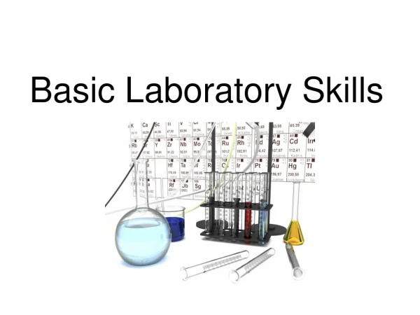
Apparatus and Equipments Dr. K. Perveen. MIC 240, K.S.U
1. Microscope • Used to observe very small organisms Dr. K. Perveen. MIC 240, K.S.U
2. Autoclave • It is a wet/ type sterilizer. • Used to sterilize culture media, glassware etc. • Usually it operates at 15 lb/sq. inch steam pressure (121.5 o C) for 30 min. Dr. K. Perveen. MIC 240, K.S.U
3. Incubator • Provide suitable temperature for the growth of organism Dr. K. Perveen. MIC 240, K.S.U
4. Hot Air Oven • It is a dry air type sterilizer. • It is used for sterilizing laboratory glass ware. • It operates at a temperature of 160 to 180 oC for one and a half hour. Dr. K. Perveen. MIC 240, K.S.U
5. Laminar Air Flow • It is a chamber which provide microbe free environment. • It is transfer of media for culturing bacteria or fungi or any microbes. Dr. K. Perveen. MIC 240, K.S.U
6. Centrifuge It spins liquid samples to separate their components Dr. K. Perveen. MIC 240, K.S.U
7. Spectrophotometer • uses wavelength to determine the concentration of a compound or particles in a solution or suspension. Dr. K. Perveen. MIC 240, K.S.U
8. Micropipette • Pipettes are used to accurately measure and dispense small volumes of liquid. Dr. K. Perveen. MIC 240, K.S.U
9.Inoculating loops & Inoculating needles • Used for inoculating microbes in the liquid media & solid media Dr. K. Perveen. MIC 240, K.S.U
10. Bunsen burner • Source of flame Dr. K. Perveen. MIC 240, K.S.U
11. Slide • Glass support for specimens Dr. K. Perveen. MIC 240, K.S.U
12.Cover Slip • Glass cover for specimens Dr. K. Perveen. MIC 240, K.S.U
13. Petri Dishes Dr. K. Perveen. MIC 240, K.S.U
14. Gram Staining Kits Dr. K. Perveen. MIC 240, K.S.U
15. Growth Media/Culture Media Dr. K. Perveen. MIC 240, K.S.U
16. Disinfectant • Clorox Bleach, diluted to 5-10% is the best cleaning agent for labs. Dr. K. Perveen. MIC 240, K.S.U
Microscopy Dr. K. Perveen. MIC 240, K.S.U
CompoundMicroscope • Instrument for observing small objects • Magnify images up to 2000X their size Dr. K. Perveen. MIC 240, K.S.U
Different parts of a microscope Dr. K. Perveen. MIC 240, K.S.U
Revolving nosepiece Eyepiece Body tube Objective Coarse adjustment Clip Fine adjustment Condenser Arm Stage Iris diaphragm Condenser control knob Mirror Base Dr. K. Perveen. MIC 240, K.S.U
Microscope Dr. K. Perveen. MIC 240, K.S.U
Resolution • It is the ability to differentiate two objects close together as being separate. Dr. K. Perveen. MIC 240, K.S.U
Magnification • Total magnification: To calculate the total magnification of any specimen being viewed multiply the power of the eye piece (ocular lens) by the power of the objective lens being used. Dr. K. Perveen. MIC 240, K.S.U
Example • If • the eye piece magnifies = 10X • the objective lens magnifies = 40X, • then 10 x 40 = 400X (total magnification) Dr. K. Perveen. MIC 240, K.S.U
Care of the microscope • Mentioned in the text Dr. K. Perveen. MIC 240, K.S.U
Types of Microscopy • Brightfield Microscope:This microscope is used to observe nonviable, stained preparation. • Darkfield Microscope • Phase-Contrast Microscope: The unstained microorganisms are observed easily. Dr. K. Perveen. MIC 240, K.S.U
Fluorescent Microscope: This microscope is used to observe the specimens that are chemically treated with a fluorescent dye. • The specimens are illuminated with an ultraviolet light. • This microscope is used for the detection of antigen-antibody reactions. • The fluorescent portion of the dye becomes visible against the black background. Dr. K. Perveen. MIC 240, K.S.U
Fluorescent image of cultured rat-brain cells. Living cells stain with calcein (left) and dead cells stain with propidium iodide (right). Dr. K. Perveen. MIC 240, K.S.U
Electron Microscope: It produces an electronically-magnified image of a specimen for detailed observation. • It is used to observe submicroscopic cellular particles as well as viral agents. • The specimen is illuminated by a beam of electrons rather than light. Dr. K. Perveen. MIC 240, K.S.U
How to Make a Wet Mount Water drop Cover slip Object/specimen Slide Dr. K. Perveen. MIC 240, K.S.U
Technique for Adding a Stain when making a Wet Mount Dr. K. Perveen. MIC 240, K.S.U
Exercise 1: Prepare the wet mount of the yeast from the culture provided to u. Observe the slide under low and high power and draw the diagram in the result sheet. Dr. K. Perveen. MIC 240, K.S.U
Observations and Results Exercise 1a Name:___________________________________________ I.D No.___________________________________________ Date:__________________ Group:________________ Dr. K. Perveen. MIC 240, K.S.U
Thanks Dr. K. Perveen. MIC 240, K.S.U