
Human physiology
E N D
Presentation Transcript
Human physiology BY: SOORYA.R XI A
CHAPTER 16DIGESTION AND ABSORPTION DIGESTION: “The breaking down of complex and insoluble organic substances such as carbohydrates, proteins and fats into simpler and soluble substances like glucose, amino acids and fatty acids respectively so that they can be easily absorbed into the body is known as digestion. This is a hydrolytic process and is carried out by various enzymes.”
ALIMENTARY OR DIGESTIVE SYSTEM:Alimentary canal is a tube present in all higher animals starting from mouth and reaching up to anus. Various glands located on its wall produce digestive juices that help in the process of digestion. Two glands namely liver and pancreas are also associated with it. They also produce the digestive juices. The digested food is also absorbed into the alimentary canal and undigested and indigestible food is passed out of the body through anus.
Mouth leads into a buccal cavity. The opening of the mouth is provided with lips. At the floor of the buccal cavity a muscular tongue is present. It helps in the ingestion, mastication and swallowing of food. It has got taste buds on its surface. Most of the mammals possess teeth on both the jaws. They are present in the cavity or socket of gums (thecodont dentition). The number and types of teeth vary in mammals. In man, there are 32 teeth of four different types namely incisors, canines, premolars and molars. This type of dentition is known as heterodont dentition. Their number can be represented by the dental formula: In each half of jaw;
Upper jaw I(2); C(1); PM(2); M(3)----------------------------------------------- = 32Lower jaw I (2); C (1); PM (2); M (3) The incisor teeth are chisel-shaped and have sharp cutting edges. Canines are dagger-shaped and pierce the food. They are very large and well developed in predatory animals. Premolars and molars are broad and strong crushing teeth. Thus the incisors are used for biting; the canines for tearing the food; and premolars and molars for grinding the food. With the help of the teeth, tongue and jaw movements, food is chewed and mixed with saliva in the mouth.
The mouth leads to a funnel-shaped pharynx, which communicates with a long muscular tube called oesophagus. The oesophagus opens into a muscular sac like structure called as stomach. In man, it is somewhat J-shaped and occupies the left side of the abdomen. The stomach opens into the small intestine. The stomach has many glands on its wall. Stomach wall produces gastric juice, which chiefly contains HCl, mucin and two protein digesting enzymes – rennin and pepsin. The muscles of the stomach wall churn and mix the food with gastric juice. Stomach through its pyloric region opens into small intestine. It is differentiated into three regions viz., duodenum, jejunum and ileum.
Small intestine is the main region where digestion and absorption of food occurs. It has large number of tubular glands that produce the intestinal juice containing a number of enzymes, which digest various types of food. Digestion of different nutrients is completed in the small intestine by the action of pancreatic juice, intestinal juice and bile juice. The end products of digestion are then absorbed from the small intestine. The small intestine opens into the large intestine. It is comparatively much shorter and wider than the small intestine. It does not have villi. It is also differentiated into three regions: caecum, colon and rectum. Caecum is a small pouch-like structure and its main part is vermiform appendix. However, caecum is very well developed in herbivorous animals like horse and ass. The colon is longest and has four parts; ascending colon, transverse colon, descending colon and pelvic colon. The pelvic colon opens into the rectum. Rectum is the last part of large intestine.
GLANDS ASSOCIATED WITH DIGESTION Pancreas: It is located in between the loops of duodenum. It is the second largest gland of the body. It secretes pancreatic juice that contains large number of digestive enzymes for digesting starch, lipids, proteins and nucleic acids. The pancreatic juice is released into the pancreatic duct, which joins with the common bile duct.
Liver: It is the largest gland of the body lying immediately below the diaphragm in the right upper part of abdomen. The cells of the liver (hepatic cells) produce bile juice that contains bile pigments and bile salts. These bile salts help in the digestion and absorption of fats. Bile juice does not contain any enzyme. Bile juice flows out of the liver through hepatic ducts forming the common bile duct that opens into the duodenum (when the food is present in the duodenum). When there is no food in the duodenum, then bile juice is stored in the gall bladder. The gall bladder is a small elongated, muscular sac below the liver. When the food comes into duodenum, it contracts to release the bile juice.
DIGESTION OF FOOD Digestion of Carbohydrates: Carbohydrates are of three types: polysaccharides, disaccharides and monosaccharides. During the process of digestion both poly-and disaccharides are broken down to monosaccharides and in this form they can be absorbed into the body. Some of these complex carbohydrates are starch and cellulose, present in cereal grains, potato, fruits and tubers; sucrose present in cane sugar; lactose present in milk etc. Enzymes that act on carbohydrates are collectively known as carbohydrases.
In the mouth cavity, the food is mixed with saliva. It contains an enzyme called salivary amylase or ptyalin. Salivary amylase acts on starch and convert it into maltose, isomaltose and small dextrins or `limit dextrin’(disaccharides). Chewing and mastication of food increases the action of salivary amylase on starch by increasing the surface area of food on which the enzyme acts. About 30 percent of starch present in food is hydrolysed in the mouth. The action of salivary amylase continues for sometime even in the stomach but soon HCl present in the gastric juice destroys the entire enzyme. Salivary Starch --------------> Maltose + Isomaltose + Dextrin Amylase Amylase Starch ------------> Maltose + Isomaltose + Dextrin Maltase Maltose + Isomaltose + Dextrin ------------> GlucoseIsomaltase
DIGESTION OF PROTEINSProteins are complex organic compounds made up of single units called amino acids. In the process of digestion, proteins are broken down to amino acids. Enzymes that hydrolyze protein are collectively known as proteases or peptides. Many of these enzymes are secreted in their inactive form or proenzymes. These inactive enzymes are converted to their active form only at the site of action.Protein digestion starts in the stomach. The gastric glands of stomach produce a light coloured, thin and transparent gastric juice. It contains hydrochloric acid and pepsinogen. The H+ ions present in HCl converts pepsinogen into pepsin. The presence of HCl makes the medium highly acidic so that pepsin can act on proteins to convert them into peptones. HCl also helps to kill bacteria and other harmful organisms that may be present along with the food. Calf gastric juice contains another milk coagulating protease, called rennin.
DIGESTION OF FATSFat digestion starts only when the food reaches the small intestine. It starts with the action of bile juice from liver. Bile juice contains bile salts, which are secreted by the liver in the bile. Bile salts break down the bigger molecules of fat globules into smaller droplets by reducing the surface tension of fat droplets. This process is known as emulsification of fats. Lipase is the enzyme that acts on emulsified fats. It is present both in the pancreatic juice and intestinal juice. Lipase converts emulsified fats into diglycerides and monoglycerides releasing fatty acids at each step. At the end of digestion, all fats are converted into fatty acids, glycerol and monoglycerides.
ABSORPTION During the process of digestion proteins are changed to amino acids, carbohydrates to glucose, fructose and galactose, fats to fatty acids, glycerol and monoglycerides. These end products of digestion are finally absorbed in small intestine. So absorption can be defined as a process by which nutrient molecules are taken into the cells of the body. Passive absorption: When the nutrients are absorbed by simple diffusion, then it is known as passive absorption. Various amino acids and monosaccharides diffuse into the blood capillaries of villi. Water is absorbed from the intestine to the intestinal cells and finally to the blood by the process of osmosis. This occurs when the solute concentration in the blood is higher (hypertonic). Thus, whenever any solute is absorbed from the intestine, it also results in the absorption of water. Active absorption: This process occurs against the concentration gradient, i.e., nutrients may be more in intestinal cells than in the lumen of intestine. It requires the expenditure of energy i.e., ATP. Various nutrients like amino acids, glucose, galactose, Na+ ions can be absorbed by active transport. After their passive absorption, they are completely absorbed by active transport.
Disorders of Digestive System:1. Jaundice: The liver is affected; skin and eyes turn yellow due to the deposit of bile pigment.2. Vomiting: It is the ejection of stomach contents through the mouth and controlled by the centre in the medulla oblongata.3. Diarrhoea: Abnormal bowel movement and the faecal discharge with more liquidity, which leads to dehydration.4. Constipation: the feces are retained within the rectum due to irregular bowel movement.5. Indigestion: food is not properly digested leading to a feeling of fullness due to inadequate enzyme secretion, anxiety, food poisoning, over eating nd spicy food.************************
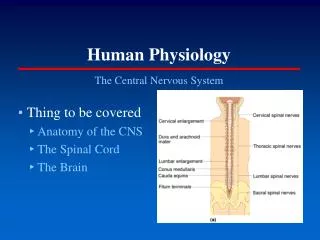
CHAPTER 17BREATHING AND EXCHANGE OF GASES • Respiration: • The process of exchange of O2 (from the atmosphere) with CO2 (produced by cells) is called breathing. This process is commonly known as respiration. • Respiratory Organs: • Mechanisms of breathing vary among different animal groups. It usually depends on the habitat and level of organization. • In case of lower invertebrates, exchange of gases takes place by simple diffusion over the entire body surface, e.g. sponges, coelenterates, flatworms, etc. • The moist cuticle of earthworms facilitates exchange of gases. • Insects have a network of tubes through which air is transported within the body. These tubes are called tracheae. • Gills are special vascularised structures which are present in most of the aquatic arthropods and mollusks. • In terrestrial animals, vascularised bags; called lungs; are present for the exchange of gases.
RESPIRATION The process of exchange of O2 (from the atmosphere) with CO2 (produced by cells) is called breathing. This process is commonly known as respiration. Respiratory Organs: Mechanisms of breathing vary among different animal groups. It usually depends on the habitat and level of organization. In case of lower invertebrates, exchange of gases takes place by simple diffusion over the entire body surface, e.g. sponges, coelenterates, flatworms, etc. The moist cuticle of earthworms facilitates exchange of gases. Insects have a network of tubes through which air is transported within the body. These tubes are called tracheae. Gills are special vascularised structures which are present in most of the aquatic arthropods and mollusks. In terrestrial animals, vascularised bags; called lungs; are present for the exchange of gases.
3. Larynx: It is a chamber situated in the region of neck. It is supported by four cartilages : thyroid is the largest and in the form of a broad ring incomplete dorsally, cricoid is a complete ring lying at the base of thyroid, a pair of arytenoids lying above the thyroid but in front of cricoid, and epiglottis situated behind the tongue that serves to cover the entrance to the trachea so that food particles may not enter into it. Larynx is also known as voice box since it helps in the production of sound. 4. Trachea: It is a tube starting from larynx running through the neck and the thoracic cavity. The trachea runs through the neck in front of the oesophagus. The trachea or windpipe is about 12 cm long and divided into two bronchi in the thoracic region.
5.Bronchi and bronchioles: The two bronchi enter into right and left lungs of either side. Inside the lungs they further branch into many smaller bronchioles with a diameter of about 1 mm. These bronchioles further divide into terminal and then into respiratory bronchioles. Each respiratory bronchiole divides into a number of alveolar ducts that further divide into atria, which swell up into air sacs or alveoli. 6. Lungs: A pair of conical shaped lungs is situated in the double walled sacs called pleural cavities. They are spongy and richly supplied with blood vessels and capillaries. They have about 300-400 millions of alveoli through which exchange of gases occur. Lungs have various bronchioles ending into alveoli where exchange of gases occurs. The alveoli are thin walled pouches the walls of which have epithelial linings supported by basement membrane.
Mechanism of breathing or pulmonary respiration:· Respiration involves the following steps:· Breathing or pulmanory ventilation by which atmospheric air is drawn in and Carbon dioxide air released out.· Diffusion of gses of oxygen and carbon dioxide across alveolar membrane.· Transport of gases by the blood.· Diffusion of oxygen and carbon dioxide between blood and tissues.· Utilisation of oxygen by the cells for catabolic reactions and release of carbon
Mechanism Of RespirationInspiration: During this process, some intercostal muscles contract thus pulling the ribs upwards and outwards. Lateral thoracic walls also move outwards and upwards. At the same time the diaphragm becomes flattened as it moves down towards the abdomen. This results in the increase in the volume of thoracic cavity thus lowering the pressure in the lungs. To fill up this gap, air from outside rushes in to bring about inhalation or inspiration. Hence, inspiration is brought about by contraction of the diaphragm and some intercostal muscles; these muscles are known as inspiratory muscles.
Expiration: During this process, the ribs return back to their original position, inwards and backwards, by the relaxation of intercostal muscles and also the diaphragm becomes dome-shaped again. Lateral thoracic walls also move inwards and downwards. This decreases the volume of the thoracic cavity thus increasing the pressure inside the lungs. So the air from the lungs rushes out through the respiratory passage bringing about expiration or exhalation.A person breathes about 12 to 16 times per minute while at rest. However, this breathing rate is higher at the time of muscular exercise and in small children.In forceful expiration, a different group of intercostal muscles and some abdominal muscles contract to reduce the volume of the thorax more than that in ordinary expiration. So more air is expelled out. Such muscles are known as expiratory muscles.
Pulmonary air volumes: Air flows into and out of the lungs because of the pressure gradient. Spirometer is an instrument used to measure the amount of air exchanged during breathing. Some terms regarding pulmonary air volumes are as follows:1. Tidal volume: It is the volume of air that is breathed in and breathed out while sitting at rest (effortless respiration) or “quiet breathing”. It is about 500 ml in an adult person. 2. Vital capacity: It is the volume of air that can be maximum expelled out after a maximum inspiratory effort. It is about 4,500 ml in males; and 3,000 ml in females. The higher the vital capacity, the greater will be the capacity for increasing the ventilation of lungs for exchange of gases. It is more in athletes and mountain dwellers. 3.Residual volume: It is the volume of air that remains inside the lungs and respiratory passage( about 1.5 litres ) after a maximum forced exhalation.4. Inspiratory reserve volume (IRV): It is the volume of air that can be taken in by forced inspiration over and above the normal inspiration or tidal volume. It is about 2,000 ml to 3,500 ml.5. Expiratory reserve volume (ERV): It is the volume of air that can still be given out by forced expiration over and above the normal inspiration or tidal volume. It is about 1,000 ml.6. Total lung capacity: It is the volume of air in the lungs and respiratory passage after a maximum inhalation effort. It is equivalent to vital capacity plus residual volume. It is about 5,000 to 6,000 ml in adult males.
Pulmonary exchange of gases:In most of higher animals including man, the air from outside reaches up to the alveoli of lungs in the process of breathing. This inspired air contains about 21 per cent oxygen, 0.04 per cent carbon dioxide, 78.6 per cent nitrogen and small amounts of other gases and atmospheric moisture. In this inspired air the partial pressure of oxygen (Po2) is 158 mm Hg; and that of carbon dioxide (Pco2) is 0.3 mm Hg. The lungs and alveoli also contain some air even after expiration. But this air has more of carbon dioxide and less of oxygen than the inspired air. So when this air mixes with the inspired air the partial pressure of oxygen in alveolar air now becomes 100 mm Hg and that of carbon dioxide becomes 40 mm Hg. However, the percentage of oxygen now becomes 13.1% and that of carbon dioxide 5.3%.
GAS TRANSPORT IN BLOODOxygen transport : The hemoglobin pigment of blood mainly transports oxygen. From alveoli of lungs, oxygen can readily diffuse into erythrocytes and combines loosely with hemoglobin (Hb) to form a reversible compound oxyhemoglobin (HbO2). Combining of oxygen with hemoglobin to form oxyhemoglobin is a physical process. There is no change in the valency of iron atom; it is ferrous in oxyhemoglobin and also in hemoglobin. This reaction, therefore, is an “oxygenation” process and not oxidation. When fully oxygenated, hemoglobin has about 97 per cent of oxygen. Hemoglobin is dark red in colour; whereas oxyhemoglobin is bright red in colour. Inside the tissues, as the partial pressure of oxygen is less, oxyhemoglobin gets dissociated into oxygen and hemoglobin. Further, as Po2 is much lower and Pco2 is much higher in active tissues than in passive tissues, so much of oxygen is released from oxyhemoglobin in active tissues. High tension of oxygen favours the formation of oxyhemoglobin while low tension of oxygen favours its dissociation. However, very little of oxygen is found in the blood plasma. Each decilitre of blood releases up to 4.6 ml, of oxygen in the tissues, 4.4 ml from oxyhemoglobin and 0.17 ml from the dissolved oxygen in the plasma.
Carbon dioxide transport :Carbon dioxide is produced in the tissues as an end product of tissue respiration. For its elimination, it gets dissolved in tissue fluid and passes into the blood. In the tissues, 100 ml of blood receives about 3.7 ml of carbon dioxide. It is transported both by the plasma and hemoglobin of blood. From the tissues, carbon dioxide diffuses into the blood plasma and forms carbonic acid (H2CO3-) in the presence of an enzyme carbonic anhydrase. Inside the erythrocytes, some of the carbonic acid forms bicarbonates and is thus transported. As carbonic acid, carbon dioxide is transported by blood plasma.
CO2 + H2O \================\ H2CO3 (carbonic acid)H2CO3 \================\ H+ + HCO3- (bicarbonate) If all the carbon dioxide produced by the tissues is carried by blood plasma in this way, then pH of the blood will be lowered to about 4.5. This would immediately cause death. So only about 10% of the CO2 produced by the tissue is actually transported as carbonic acid. About 20% of the total CO2 produced is transported by the hemoglobin of blood as carbaminohemoglobin. CO2 + Hb.NH2 ---------------------à Hb.NH.COOH
Gas exchange in tissues: In the tissues, gases are exchanged by diffusion (as in the lungs). In tissues, as the partial pressure of oxygen is very low (about 40 mm Hg), so the oxygen gets unloaded here. When the blood leaves the tissues it has Po2 of 40 mm Hg. However, for carbon dioxide it is just the reverse. The blood entering into tissues has Pco2 of 40 mm Hg; while Pco2 of tissues is 46 mm Hg. So some of carbon dioxide from tissues gets loaded into the blood. Disorders of Respiratory System:1. Asthma: difficulty in breathing causing wheezing due to inflammation of bronchi and bronchioles.2. Emphysema: alveolar walls are damaged due to which respiratory surface is decreased, causes of this is cigarette smoking.3. Occupational Respiratory Disorders: long exposure to the dust of industries like stone breaking, etc cause an inflammation on lung tissues and leads to lung damage.
Distinguish between:1. Inspiratory muscles and expiratory muscles : Inspiratory muscles are a group of intercoastalmuscles, the contraction and relaxation of which bring about inspiration and expiration respectively. Expiratory muscles are a group of different intercoastalmuscles and some abdominal muscles which contract to reduce the thoracic cavity more than that in ordinary expiration as in forceful respiration.2. Tracheaolesand bronchioles :Tracheaolesare the finer branches of tracheal tubes present in insects that ramify into the tissues. Bronchioles are the finer branches of bronchus that branch further to open into alveoli of lungs in mammals.3. Carbamino-hemoglobin and oxy-hemoglobin :Carbamino-hemoglobin is a reversible compound formed when hemoglobin combines with carbon dioxide; oxy-hemoglobin is a reversible compound formed when hemoglobin combines with oxygen. In carbon monoxide poisoning, Hb combines irreversibly with CO to form carboxyhemoglobin.
1. Far more oxygen is released from oxyhemoglobin in a more active tissue than in a less active one.The dissociation of oxyhemoglobin to oxygen and deoxyhemoglobin depends upon the partial pressures of oxygen and carbon dioxide in the tissues. In a more active tissue, the Po2 is lower and Pco2 is higher as compared to that of a less active tissue. So far more oxygen is released from oxyhemoglobin in a more active tissue than in a less active one. 2. Oxygenation of blood promotes the release of carbon dioxide from the blood in the lungs.The oxygenation of blood in the lungs depends on the partial pressures of oxygen in the pulmonary artery and in the alveolar air. Further, the oxygen affinity of hemoglobin is enhanced with the fall in partial pressures of carbon dioxide that results from the elimination of carbon dioxide from the blood into the lungs. In the lung alveoli, hemoglobin is exposed to high Po2 and less Pco2. 3. Oxygen leaves the blood from tissue capillaries, but carbon dioxide enters the blood in tissue capillaries.Inside the tissue capillaries, there is more of Pco2 and less of Po2. The blood coming to tissues has more of Po2 and less of Pco2. So oxygen is unloaded from the blood and carbon dioxide is loaded to the blood in the tissue capillaries.
4. Erythrocytes can carry out anaerobic metabolism only.Erythrocytes can carry out anaerobic metabolism only because they lack mitochondria.5. Gaseous exchanges continue in the lungs without interruption during expiration. Gaseous exchanges continue in the lungs uninterrupted because some air is always present inside the lung alveoli even during expiration. 6. Contraction of inspiratory muscles causes inspiration while relaxation causes expiration.Contraction of inspiratory muscles increases the volume of pleural cavities; while expiration is brought about passively by the relaxation of those muscles. As the muscles relax, the diaphragm moves up towards the thorax and the intercostal muscles move the lateral thoracic walls inwards and downwards. This decreases the volume of pleural cavities and the air rushes out. 7. Oxygen enters the blood from the alveolar air but carbon dioxide leaves the blood to enter the alveolar air.Inside the alveoli of lungs, there is more of Po2 and less of Pco2 .The blood coming to lung alveoli has more of Pco2 and less of Po2. So oxygen is loaded to the blood and carbon dioxide is unloaded from the blood in the alveoli of lungs.