Download

1 / 75
770 likes | 1.53k Vues
SPECT IN CONCUSSION, VERTEBRO-BASILAR INSUFFICIENCY AND OTHER NEUROLOGICAL DISORDERS. Novel applications and insights. Deepak Agrawal, MBBS, MS, MCh. Fellow, Pediatric Neurosurgery BC Children’s Hospital, UBC. LEARNING OBJECTIVES.

E N D
SPECT IN CONCUSSION, VERTEBRO-BASILAR INSUFFICIENCY AND OTHER NEUROLOGICAL DISORDERS Novel applications and insights • Deepak Agrawal, MBBS, MS, MCh. • Fellow, Pediatric Neurosurgery • BC Children’s Hospital, UBC
LEARNING OBJECTIVES • To understand uses of Single Photon Emission Computed Tomography (SPECT) in patients with post-concussion syndrome and vertebrobasilar insufficiency • To be familiar with the use of SPECT as an investigative and research tool in neurosciences
Imaging Brain function using SPECT All India Institute of Medical Sciences, New Delhi
Imaging Brain function using SPECT All India Institute of Medical Sciences, Delhi • 1200 bed tertiary centre • Annual budget of CAD $250 million Imaging • 6 CT scanners • 2 1.5 T MRI scanners, a 4.7 Tesla animal MRI/MRS scanner, and a 9.4 Tesla NMR spectrometer. • 1 PET, 2 SPECT scanners
Imaging Brain function using SPECT IMAGING TECHNIQUES • STRUCTURAL- MRI, CT • FUNCTIONAL- SPECT, PET, fMRI (SPECT-Single Photon Emission Computed Tomography)
Imaging Brain function using SPECT Functional Imaging • PET- Gold standard • SPECT - More widely available - Much cheaper - Still able to provide much of the same information
Imaging Brain function using SPECT UNDERLYING PRINCIPLES OF SPECT • Biochemical interactions between brain tissue and injected substance (HMPAO, ECD, Iophane) • These substances are labeled with radioactive tracers (99mTc-HMPAO, 99mTc-ECD, 123I-iophane)
Imaging Brain function using SPECT UNDERLYING PRINCIPLES • Radiopharmaceutical injected IV • Crosses BBB proportionate to blood flow • Enters Neuronal tissue – stereoisomeric change • Become ‘trapped’ inside the neurons
Imaging Brain function using SPECT UNDERLYING PRINCIPLES • Increased neuronal activity = increased neuronal uptake of radiopharmaceutical = Increased perfusion on SPECT • This image of cerebral perfusion indirectly reflects cerebral metabolism
Imaging Brain function using SPECT BRAIN SPECT • Brain perfusion - HMPAO,ECD HMPAO-Hexa Methyl Propylene Amine Oxine; ECD-Ethylene Cysteinate Dimer • Imaging of neurotransmitter systems - Ioflupane
Imaging Brain function using SPECT APPLICATIONS • Dementias Alzheimers:↓perfusion TP regions B/L Normal perfusion Subcortical region Vascular Dementia:↓perfusion Subcortical regions
Imaging Brain function using SPECT APPLICATIONS • Epilepsy Established role in epilepsy SISCOM(Subtraction Ictal SPECT Coregistered with MRI)
Imaging Brain function using SPECT APPLICATIONS • Trauma Can show abnormalities in pts with normal CT & MRI Sensitivity can reach 80% compared to 5% for CT and 60% for MRI in minor head injuries Kant R, Smith-Seemiller L, Isaac G, Duffy J. Tc-HMPAO SPECT in persistent post-concussion syndrome after mild head injury: comparison with MRI/CT. Brain Inj 1997;11:115.
Imaging Brain function using SPECT APPLICATIONS • Parkinsonism Dopamine transporter imaging- assessing the presynaptic dopaminergic function
Imaging Brain function using SPECT APPLICATIONS • Research tool before & after pharmacotherapy, psychotherapy & surgery
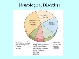
Imaging Brain function using SPECT APPLICATIONS • Dementias • Epilepsy • Trauma • Parkinsonism • Stroke • Research tool-(before & after pharmacotherapy, psychotherapy, surgery)
Imaging Brain function using SPECT APPLICATIONS • Dementias • Epilepsy • Trauma • Parkinsonism • Stroke • Research tool-(before & after pharmacotherapy, psychotherapy, surgery)
Imaging Brain function using SPECT TRAUMA • 90% of all HI are supposedly ‘minor’ • Post concussion syndrome—including symptoms such as headache, irritability, poor concentration, memory disturbances, dizziness, anxiety, and depression—has been reported to occur in up to 80% of the patients following minor HI. Hugenholtz H, Stuss DT, Stethem LL, Richard MT: How long does it take to recover from a mild concussion? Neurosurgery 22:853–858, 1988 Wood RL. Understanding the 'miserable minority': a diasthesis-stress paradigm for post-concussional syndrome. Brain Inj.2004;18(11):1135-53
Medial Temporal Injury In Pediatric Postconcussion Syndrome • Deepak Agrawal, Naveen K*, C S Bal*, A K Mahapatra • Departments of Neurosurgery and *Nuclear medicine, All India Institute of Medical Sciences, New Delhi
Temporal lobe damage in minor head injury • WHY MEDIAL TEMPORAL LOBE? • Hippocampus is especially vulnerable to insults such as ischemia, hypoxia, and seizures • Extent of hippocampal damage can be correlated with severity of memory impairment Rempel-Clower NL, Zola SM, Squire LR, Amaral DG. Three cases of enduring memory impairment after bilateral damage limited to the hippocampal formation. J Neurosci 1996;16:5233-5255.
Temporal lobe damage in minor head injury OBJECTIVES • Look for medial temporal hypoperfusion (MTH) on SPECT in children with minor head injury • To evaluate MTH on SPECT as a risk factor for development of persistent postconcussion syndrome (PPCS) at three months
Temporal lobe damage in minor head injury MATERIALS AND METHODS • PROSPECTIVE STUDY • One year period • Children ≤ 18 yrs of age
Temporal lobe damage in minor head injury MINOR HEAD INJURY • Loss of consciousness <30 minutes. • GCS score 13 to 15. • Posttraumatic amnesia <24 hours. [criteria published by the members of the Mild Traumatic brain injury Interdisciplinary Special Interest Group (BISIG)] Kay T, Harrington DE, et al. Definition of mild traumatic brain injury. J Head Trauma Rehabil 1993;8:86
Temporal lobe damage in minor head injury POST CONCUSSION SYNDROME: (2 or more of the following) • Headache • Dizziness and vertigo • Memory deficits • Behavioral and emotional disturbances. DSM IV criteria Brown SJ, Fann JR, Grant I: Postconcussion disorder: time to acknowledge a common source of neurobehavioural morbidity. J Neuropsychiatry Clin Neurosci 6:15-22, 1994
Temporal lobe damage in minor head injury • CT head INVESTIGATIONS • SPECT scan brain (Within 72 hours & at 3 months)
Temporal lobe damage in minor head injury SPECT METHODOLOGY • SPECT scanning was done using 99Tcm-ECD on a dual headed GE 'Varicam' scanner. • The final data was displayed on a 10 grade color scale and semi quantitative analysis performed.
Temporal lobe damage in minor head injury STUDY DESIGN
Temporal lobe damage in minor head injury RESULTS (SPECT) • 13/14 (93%) patients with initial MTH continued to have persistent MTH • 0/16 (0%) of patients in control group developed subsequent MTH at 3 months
Baseline At 3 months
Temporal lobe damage in minor head injury RESULTS Persistent post concussion syndrome (PPCS) 12/14 (86%) children developed PPCS in the MTH group, compared to 2/16 (12.5%) children in the control group P=0.0003
Temporal lobe damage in minor head injury CONCLUSIONS • Children with MTH are more likely to develop persistent post concussion syndrome • SPECT may help in identification and prognostication in this subgroup of children
ROLE OF PIRACETAM IN POST-CONCUSSION SYNDROME A PROSPECTIVE RANDOMIZED STUDY Deepak Agrawal, K Naveen*, CS Bal*, AK Mahapatra Departments of Neurosurgery* and Nuclear medicine*, ALL INDIA INSTITUTE OF MEDICAL SCIENCES, NEW DELHI, INDIA
Piracetam in minor head injury Piracetam • Discovered 30 years ago by UCB pharma • GABA derivative, does not work via GABA pathways. • Mechanism of action remains unknown • Has beneficial effects on the CBF by decreasing the adhesivity, aggregation, and increasing the deformability of erythrocytes. • “Nootropic”
Piracetam in minor head injury Why Piracetam? • Already approved for use in Europe & India for a variety of disorders including head injury • Minimal side effects- More than 12 g of piracetam has been given as daily dose in acute stroke without appreciable side effects De Deyn PP, Reuck JD, Deberdt W, Vlietinck R, Orgogozo JM. Treatment of acute ischemic stroke with piracetam. Members of the Piracetam in Acute Stroke Study (PASS) Group. Stroke 1997; 28: 2347-52.
Piracetam in minor head injury • OBJECTIVE To look for changes in cerebral perfusion on SPECT, following administration of Piracetam, in patients with post-concussion syndrome (PCS),
Piracetam in minor head injury MINOR HEAD INJURY 1. Loss of consciousness <30 minutes. 2. GCS score 13 to 15. 3. Posttraumatic amnesia <24 hours. [criteria published by the Mild Traumatic brain injury Interdisciplinary Special Interest Group (BISIG)] Kay T, Harrington DE, et al. Definition of mild traumatic brain injury. J Head Trauma Rehabil 1993;8:86
Piracetam in minor head injury • Headache • Dizziness and vertigo • Memory deficits • Behavioral and emotional disturbances. POST CONCUSSION SYNDROME: (2 or more of the following) DSM IV criteria based on recommendation of Brown et al. Brown SJ, Fann JR, Grant I: Postconcussion disorder: time to acknowledge a common source of neurobehavioural morbidity. J Neuropsychiatry Clin Neurosci 6:15-22, 1994
Piracetam in minor head injury MATERIALS AND METHODS • Prospective study • Adult patients (18-60 yrs)
Piracetam in minor head injury • CT head INVESTIGATION • SPECT scan brain Within 72 hours of injury
Piracetam in minor head injury Circular region of interest (ROI) with a radius of 9.1mm (6 pels) in the basal ganglia, thalamus, temporal lobe, visual cortex and brain stem were used. SPECT
Piracetam in minor head injury Semiquantitative analysis of the data done using semiautomatic brain quantification programs (Xpertpro, Entegra with Neurogam). ABNORMAL SPECT SCAN Regional cerebral perfusion <10% of contralateral lobe, or in case of bilateral involvement, less than 20% of cerebellum
Piracetam in minor head injury STUDY DESIGN
RESULTS • Significant rise in the post treatment ratio in the piracetam group (mean: 0.959) as compared to the controls (mean: 0.882) (p= <.001) • Nine patients (90%) also had improvement in their symptoms of PCS, compared to only three patients in the control group (p=0.01).
Piracetam in minor head injury CONCLUSIONS • Cerebral perfusion defects occur in majority of the cases of postconcussion syndrome following minor head injury. • Piracetam reverses cerebral perfusion deficits and may result in accelerated symptomatic improvement in patients with postconcussion syndrome.
Piracetam in minor head injury THIS PRELIMINARY STUDY SHOWED OBJECTIVELY THAT LOW DOSE PIRACETAM MAY BENEFIT PATIENTS WITH POSTCONCUSSION SYNDROME CONCLUSIONS