Download
1 / 29
300 likes | 368 Vues
Learn about cell division in different life stages: growth, repair, and reproduction. Explore prokaryotic and eukaryotic cell division, genes, homologous chromosomes, and more. Understand karyotypes, trisomy, and mutations in chromosome structure.

E N D
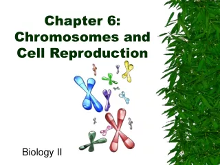
Chapter 6: Chromosomes and Cell Reproduction Biology II
Cell Division • Cell division occurs at different times in an organism’s life • Growth and development • Repair • Reproduction • Gametes – reproductive cells (egg/sperm) • When a cell divides, DNA is first copied and then distributed so that each cell ends up with a copy of DNA
Prokaryotic Cell Division • Prokaryotes have single circular DNA molecule • Reproduce by binary fission • Form of asexual reproduction that produces identical offspring • Occurs in 2 stages • DNA is copied • Cell divides by adding a new cell membrane to a point on membrane between the 2 DNA copies
Genes • Information encoded in DNA is organized into units called genes • Segment of DNA that codes for a protein or RNA molecule • Single DNA molecule has 1000s of genes linked together • Genes play important role in determining how an organism’s body develops and functions • When genes are being used, the DNA is stretched out so that its information can be used to direct protein production
Eukaryotic Cell Division • As eukaryotic cell prepares to divide, chromosomes become visible • DNA and proteins associated with DNA • Before DNA coils, DNA is copied, forming chromatids • 2 exact copies of DNA that make up each chromosome • Attached at a point called a centromere • Chromatids separate during cell division and are placed into each new cell, ensuring that each new cell will have same genetic information as the original cell
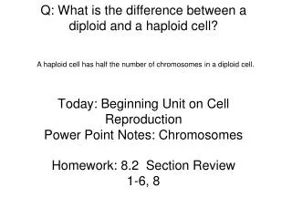
Homologous Chromosomes • Each human somatic (body) cell has 2 copies of 23 chromosomes (total of 46 chromosomes) • Each pair is made up of homologous chromosomes • Similar in size, shape, and genetic content • Each homologue comes from one of the 2 parents • Differ in size, shape, and sets of genes • Each contains 1000s of genes
Sets of Chromosomes • All cells in the body, other than gametes, are somatic cells • Said to be diploid (2n), since they contain 2 sets of chromosomes • Symbol “n” is used to represent 1 set of chromosomes (human diploid #: 2n = 46) • Gametes are said to be haploid (n), since they contain only one set of chromosomes • Fusion of 2 haploid gametes, known as fertilization, forms a diploid zygote • Fertilized egg cell
Numbers of Chromosomes • Each organism has a characteristic number of chromosomes • Number of chromosomes remain constant within each species
Sex Chromosomes • Humans have 22 pairs of autosomes and 1 pair of sex chromosomes • Autosomes – chromosomes that are not directly involved in determine the gender of an individual • Sex chromosomes – contain genes that will determine sex of individual • Referred to as X and Y chromosomes in humans and many other organisms • Sex of offspring is determined by male • XX: Female • XY: Male • In some insects (grasshoppers), females are XX and males are XO (O = absence of chromosome) • In birds, moths, and butterflies, male is XX and female is XO
Karyotypes • Presence of normal number of chromosomes is necessary for normal development and function • Abnormalities in chromosome number can be detected by analyzing a karyotype • Photo of the chromosomes in a dividing cell that shows the chromosomes arranged by size
Trisomy and Nondisjunction • Trisomy is a condition in which humans have more than 2 copies of a chromosomes • Occurs if one or more chromosomes fail to separate properly, an event known as nondisjunction • Trisomy 21 is known as Down’s syndrome • Characterized by short stature, round face with upper eyelids that cover inner corners of eyes, and varying degrees of mental retardation • More likely in pregnancies of older women because eggs can accumulate increasing amount of damage over time
Change in Chromosome Structure • Changes in organism’s chromosome structure are called mutations • Breakage of chromosomes can lead to 4 types of mutations • Deletion mutation – piece of chromosome breaks off completely • Duplication mutation – chromosome fragment attaches to its homologous chromosome • Inversion mutation – chromosome piece reattaches to original chromosome but in reverse orientation • Translocation mutation – chromosome piece reattaches to a nonhomologous chromosome
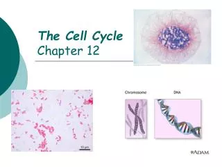
Section 6-2 The Cell Cycle
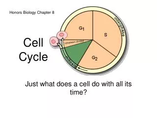
The Cell Cycle • Cell division more complicated in eukaryotes because it involves dividing chromosomes, cytoplasm, and other organelles • Cell cycle: repeating sequence of cellular growth and division during life of an organism • 90% of cell’s time is spent in first of 3 phases, collectively called interphase • Cell enters last 2 phases only if it is about to divide
Stages of Interphase • First growth phase (G1): cell grows rapidly and carries out routine functions • Cells remain in this phase until they prepare to divide • Some cells (nerve/muscle cells) never divide • Synthesis phase (S): cell’s DNA is copied • At end of this phase, each chromosome consists of 2 chromatids attached at a centromere • Second growth phase (G2): preparations are made for nucleus to divide and microtubules are rearranged in preparation for mitosis
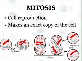
Stages of Cell Division • Mitosis: process during cell division in which nucleus of cell is divided into 2 nuclei • Each nucleus ends up with the same number and kinds of chromosomes as original cell • Cytokinesis: process during cell division in which cytoplasm divides • Recall: Mitosis and cytokinesis produce new cells identical to the original cells • Allows organisms to grow, replace damaged tissues, or to reproduce asexually
Control of Cell Cycle • Cells know when to divide based on a series of three main checkpoints at which feedback signals from cell can trigger or delay the next phase • Cell growth (G1) checkpoint: makes decision whether cell will divide • Are conditions favorable for division? • Is cell healthy and large enough? • If so, certain proteins will stimulate cell to begin synthesis phase • DNA synthesis (G2) checkpoint: DNA replication is checked by DNA repair enzymes • Mitosis checkpoint: triggers exit from mitosis and signals beginning of G1 phase
Cancer • Specific genes contain information to make proteins that regulate cell growth and division • If one of these genes is mutated, protein may not function and regulation of cell growth and division may be disrupted • Cancer: uncontrolled growth of cells • Some mutations cause cancer by overproducing growth-promoting molecules, speeding up cell cycle • Others cause cancer by inactivating control proteins that normally act to slow or stop cell cycle
Section 6-3 Mitosis and Cytokinesis
Mitosis and the Spindle • During mitosis, the chromatids of each chromosome are physically moved to opposite sides of dividing cell • Structures called spindles are involved in moving these chromosomes • Made up of centrioles and individual microtubule fibers
Spindle Formation • Organelles that organize the assembly of the spindle are called centrosomes • Found at each of the cell’s poles • In animals, a pair of centrioles is found inside each centrosome • Like spindle fibers, centrioles are made of microtubules • Each spindle fiber consists of an individual microtubule • Each centriole is made of nine triplets of microtubules arranged in a circle • Though plant cells do not have centrioles, they form a spindle almost identical to that of an animal cell
Separation of Chromatids • As cell prepares to divide, microtubules in spindle extend out toward opposite poles of cell • Once microtubules attach to centromeres and poles, the 2 chromatids (now called chromosomes) can be separated • As the paired chromatids separate, they move toward opposite poles along paths described by microtubules to which they are attached • Chromatids draw closer to poles of cell as spindle microtubules break down and become shorter • Once breakdown of the spindle is complete, each pole has one complete set of chromosomes
Stage 1: Prophase • Stage 1 of mitosis is called prophase • During this stage: • Chromosomes coil up, becoming visible • Nuclear envelope dissolves • Spindle forms
Stage 2: Metaphase • The second stage of mitosis is called metaphase • During this stage: • Chromosomes move to center of cell and line up along equator • Spindle fibers link chromatids of each chromosome to opposite poles
Stage 3: Anaphase • The third stage of mitosis is called anaphase. • During this stage: • Centromeres divide • Separated chromatids (now called chromosomes) move toward opposite poles as spindle fibers attached to them shorten
Stage 4: Telophase • The last stage of mitosis is called telophase. • During this stage: • Nuclear envelope forms around chromosomes at each pole • Chromosomes (now at opposite poles) uncoil • Spindle dissolves as spindle fibers break down and disappear
Cytokinesis • As mitosis ends, cytokinesis begins. • Cytoplasm of cell is divided in half • Cell membrane grows to enclose each cell, resulting in formation of 2 separate but genetically identical cells
Cytokinesis: Plant vs. Animal Cells • Cytokinesis varies according to cell type: • In animals (lack cell walls), cells are pinched in half by belt of protein threads • In plants (have cell walls), vesicles formed by Golgi apparatus fuse at midline of dividing cell to form a cell plate • A new cell wall then forms on both sides of cell plate, separating the plant cell into 2 genetically identical cells