Download
1 / 23
260 likes | 721 Vues
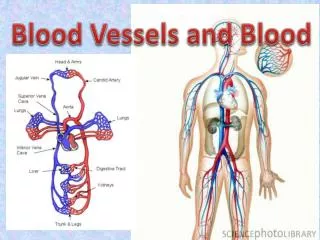
8. HEART AND BLOOD VESSELS. Major Arteries and Veins. Figure 8.10. Blood Vessels. Arterial system Structure: endothelium, middle, outer layers Functions: Arteries: carry blood away from heart Arterioles and precapillary sphincters control pressure

E N D
8 HEART AND BLOOD VESSELS
Major Arteries and Veins Figure 8.10
Blood Vessels • Arterial system • Structure: endothelium, middle, outer layers • Functions: • Arteries: carry blood away from heart • Arterioles and precapillary sphincters control pressure • Capillaries: exchange of nutrients, waste, and defensive cells between vessel and tissue
Blood Vessels (cont.) • Venous system • Structure: veins: three layers, thin-walled • Functions: carry blood toward the heart • Mechanisms in blood return: • Contraction of skeletal muscles • One-way valves • Pressure changes associated with breathing
Arterioles and Capillaries Figure 8.2
Capillary Structure Figure 8.4
Lymphatic System • Function: maintain blood volume; also functions in immune system • Structure: • Blind-ended capillaries • Lymphatic vessels • Lymph
The Heart Figure 8.8
The Heart • Structure • Layers: epicardium, myocardium, endocardium • Chambers: two atrias, two ventricles • Valves: • Two atrioventricular valves: tricuspid and bicuspid (mitral) • Two semilunar valves: pulmonary and aortic
Pulmonary Circuit: Oxygenation of Blood • Pathway: • Deoxygenated blood through the vena cava to the right atrium • Deoxygenated blood through the right atrioventricular valve to the right ventricle • Deoxygenated blood through the pulmonary semilunar valve to the pulmonary trunk and the lungs • Oxygenated blood through the pulmonary veins to the left atrium • Oxygenated blood through the left atrioventricular valve to the left ventricle
Systemic Circuit: Delivery of Oxygenated Blood to Tissues and Return of Blood to the Heart • Pathway: • Oxygenated blood through the aortic semilunar valve to the aorta • Oxygenated blood through branching arteries and arterioles to the tissues • Oxygenated blood through the arterioles to capillaries • Deoxygenated blood from capillaries into venules and veins • Ultimately to the vena cava and into the right atrium
Cardiac Cycle Figure 8.12
Heart Sounds and Heart Valves • Lub-dub • Heart murmurs
Cardiac Conduction System Coordinates Contraction • SA node: cardiac pacemaker • AV node: relay impulse • AV bundle and Purkinje fibers: carry impulse to ventricles Figure 8.14
Electrocardiograms (EKG/ECG) • Three formations: • P wave: impulse across atria • QRS complex: spread of impulse down septum, around ventricles in Purkinje fibers • T wave: end of electrical activity in ventricles • Arrythmias, ventricular fibrillation
Electrocardiograms (EKG/ECG) (cont.) Figure 8.15b,c
Blood Pressure • Definitions: “normal”: • Systolic pressure • Diastolic pressure • Measurement: sphygmomanometer
Blood Pressure (cont.) • Hypertension: high blood pressure: • Definition • The silent killer • Risk factors • Hypotension: blood pressure too low: • Clinical signs: dizziness, fainting • Causes: orthostatic, severe burns, blood loss
Regulation of the Cardiovascular System: Baroreceptors • Baroreceptors: pressure receptors in aorta and carotid arteries • Steps in mechanism: • Blood pressure rises, vessels stretched • Signals sent to brain in the cardiovascular center • Heart signaled to lower heart rate and force of contraction • Arterioles vasodilate, increasing blood flow to tissues • Combined effect lowers blood pressure
Regulation of the Cardiovascular System: Nervous and Endocrine Factors • Medulla oblongata signals: • Sympathetic nerves: constrict blood vessels, raising blood pressure • Parasympathetic nerves: dilate blood vessels, lowering blood pressure • Hormones: epinephrine (adrenaline) • Local requirements dictate local blood flow
Cardiovascular Disorders • Angina pectoris: a warning • Myocardial infarction/heart attack: permanent cardiac damage • Congestive heart failure: decrease in pumping efficiency • Embolism: blockage of blood vessels • Stroke: impaired blood flow to the brain
Reducing the Risk of Cardiovascular Disease • Smoking: don’t • Blood lipids: monitor cholesterol levels • Exercise: regular and moderate • Blood pressure: treat hypertension • Weight: being overweight increases risk of heart attack and stroke • Control of diabetes mellitus: early diagnosis and treatment delays onset of related problems • Stress: avoid chronic stress
Cardiac Anatomy Quiz Test Yourself, page 187