Download
1 / 28
351 likes | 816 Vues
Functional Organization of Nervous Tissue. The Nervous System. Components Brain, spinal cord, nerves, sensory receptors Responsible for Sensory perceptions, mental activities, stimulating muscle movements, secretions of many glands Subdivisions Central nervous system ( CNS )

E N D
The Nervous System • Components • Brain, spinal cord, nerves, sensory receptors • Responsible for • Sensory perceptions, mental activities, stimulating muscle movements, secretions of many glands • Subdivisions • Central nervous system (CNS) • Peripheral nervous system (PNS)
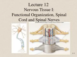
Central Nervous System • Consists of • Brain • Located in cranial vault of skull • Spinal cord • Located in vertebral canal • Brain and spinal cord • Continuous with each other at foramen magnum
Peripheral Nervous System • Two subcategories • Sensory or afferent • Motor or efferent • Divisions • Somatic nervous system • Autonomic nervous system (ANS) • Sympathetic • Parasympathetic • Enteric
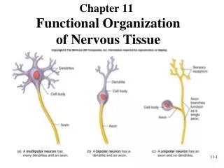
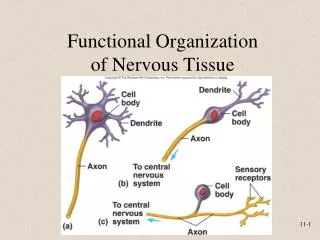
Cells of Nervous System • Neurons or nerve cells • Receive stimuli and transmit action potentials • Organization • Cell body or soma • Dendrites: Input • Axons: Output • Neuroglia or glial cells • Support and protect neurons
Types of Neurons • Functional classification • Sensory or afferent: Action potentials toward CNS • Motor or efferent: Action potentials away from CNS • Interneurons or association neurons: Within CNS from one neuron to another • Structural classification • Multipolar, bipolar, unipolar
Neuroglia of CNS • Astrocytes • Regulate extracellular brain fluid composition • Promote tight junctions to form blood-brain barrier • Ependymal Cells • Line brain ventricles and spinal cord central canal • Help form choroid plexuses that secrete CSF
Neuroglia of CNS • Microglia • Specialized macrophages • Oligodendrocytes • Form myelin sheaths if surround axon
Neuroglia of PNS • Schwann cells or neurolemmocytes • Wrap around portion of only one axon to form myelin sheath • Satellite cells • Surround neuron cell bodies in ganglia, provide support and nutrients
Myelinated and Unmyelinated Axons • Myelinated axons • Myelin protects and insulates axons from one another • Not continuous • Nodes of Ranvier • Unmyelinated axons
Electrical Signals • Cells produce electrical signals called action potentials • Transfer of information from one part of body to another • Electrical properties result from ionic concentration differences across plasma membrane and permeability of membrane
Ion Channels • Nongated or leak channels • Always open and responsible for permeability • Specific for one type of ion although not absolute • Gated ion channels • Ligand-gated • Open or close in response to ligand binding to receptor as ACh • Voltage-gated • Open or close in response to small voltage changes
Resting Membrane Potential • Characteristics • Number of charged molecules and ions inside and outside cell nearly equal • Concentration of K+ higher inside than outside cell, Na+ higher outside than inside • At equilibrium there is very little movement of K+ or other ions across plasma membrane
Changes in Resting Membrane Potential • K+ concentration gradient alterations • K+ membrane permeability changes • Depolarization or hyperpolarization: Potential difference across membrane becomes smaller or less polar • Hyperpolarization: Potential difference becomes greater or more polar • Na+ membrane permeability changes • Changes in Extracellular Ca2+ concentrations
Local Potentials • Result from • Ligands binding to receptors • Changes in charge across membrane • Mechanical stimulation • Temperature or changes • Spontaneous change in permeability • Graded • Magnitude varies from small to large depending on stimulus strength or frequency • Can summate or add onto each other
Action Potentials • Series of permeability changes when a local potential causes depolarization of membrane • Phases • Depolarization • More positive • Repolarization • More negative • All-or-none principle • Camera flash system
Refractory Period • Sensitivity of area to further stimulation decreases for a time • Parts • Absolute • Complete insensitivity exists to another stimulus • From beginning of action potential until near end of repolarization • Relative • A stronger-than-threshold stimulus can initiate another action potential
Action Potential Frequency • Number of potentials produced per unit of time to a stimulus • Threshold stimulus • Cause an action potential • Maximal stimulus • Submaximal stimulus • Supramaximal stimulus Inser
The Synapse • Junction between two cells • Site where action potentials in one cell cause action potentials in another cell • Types • Presynaptic • Postsynaptic
Chemical Synapses • Components • Presynaptic terminal • Synaptic cleft • Postsynaptic membrane • Neurotransmitters released by action potentials in presynaptic terminal • Synaptic vesicles • Diffusion • Postsynaptic membrane • Neurotransmitter removal