Download
1 / 69
710 likes | 929 Vues
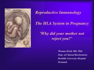
Reproductive Immunology. The HLA System in Pregnancy. ’Why did your mother not reject you?’. Thomas Hviid, MD, PhD Dept. of Clinical Biochemistry Roskilde University Hospital Denmark. Topics. Introduction: The semi-allogenic fetus Fundamental aspects of MHC/HLA and HLA class Ib

E N D
Reproductive Immunology The HLA System in Pregnancy ’Why did your mother not reject you?’ Thomas Hviid, MD, PhD Dept. of Clinical Biochemistry Roskilde University Hospital Denmark
Topics • Introduction: The semi-allogenic fetus • Fundamental aspects of MHC/HLA and HLA class Ib • ’Classical’ vs ’non-classical’ MHC/HLA genes • Gene structure, polymorphisms, expression, alternative splicing, functions etc • Certain complications of pregnancy in relation to HLA/HLA-G • (Recurrent) spontaneous abortions, IVF, pre-eclampsia • Pregnancy and HLA diversity • Reproductive selection mechanisms (’mating preferences’) and HLA
Medawar & Billingham, Nature, 1953 • Four hypotheses: • The conceptus lacks immunogenicity • Significant lowering of the immune response during pregnancy • The uterus is an immunoprivileged site • Immune barrier elaborated by the placenta: • Tolerance to the semi-allognic fetus by the maternal immune system seems mainly an active mechanism: • Fetal tissue prevented from being recognized as foreign tissue and/or being rejected by the maternal immune system
Hypotheses/concepts to explain maternal tolerance of the fetus • HLA/HLA-G expression by the trophoblast • The Th1/Th2 balance • Regulatory CD4+CD25+ T cells • Others • Leukemia inhibitory factor (LIF) • Indoleamine 2,3-dioxygenase (IDO) • Suppressor macrophages • Hormones • CD95 and its ligand • Annexin II • Lowered complement activity • Hidden trophoblast antigens (Thellin et al, review 2000)
Human Leucocyte Antigen (HLA)Major Histocompatibility Complex (MHC) • Several classes of HLA genes: • HLA class Ia (classical HLA class I antigens; on almost all cells) • HLA-A, HLA-B, HLA-C • HLA class Ib (non-classical HLA class I antigens) • HLA-E, HLA-G, HLA-F • HLA class II (expressed on antigen presenting cells, B cells) • HLA-DR, HLA-DP, HLA-DQ • Discovered / rejection of transplants (Jean Dausset 1952/1953) • The function of HLA / MHC was elucidated in the 1970s(Zinkernagel & Doherty 1974) • MHC/HLA molecules present antigen peptides to T cells via the T cell receptor • Antigen peptides (eg pathogenes) are recognized in combination with an individual’s own variant of HLA
The classical HLA class Ia molecules are highly polymorphic HLA-A10 HLA-B12 HLA-Cw5 HLA-A3 HLA-B5 HLA-Cw7 HLA-A23 HLA-B12 HLA-Cw1 HLA-A11 HLA-B16 HLA-Cw8 HLA-A25 HLA-B40 HLA-Cw2 HLA-A26 HLA-B8 HLA-Cw5 HLA-A2 HLA-B27 HLA-Cw6 HLA-A28 HLA-B17 HLA-Cw5 HLA-A19 HLA-B14 HLA-Cw8 HLA-A19 HLA-B15 HLA-Cw2 HLA-A25 HLA-B12 HLA-Cw1 HLA-A24 HLA-B8 HLA-Cw4
The non-classical HLA class Ib molecules are nearly monomorphic HLA-G HLA-E HLA-F HLA-G HLA-E HLA-F HLA-G HLA-E HLA-F HLA-G HLA-E HLA-F HLA-G HLA-E HLA-F HLA-G HLA-E HLA-F HLA-G HLA-E HLA-F HLA-G HLA-E HLA-F HLA-G HLA-E HLA-F HLA-G HLA-E HLA-F HLA-G HLA-E HLA-F HLA-G HLA-E HLA-F
HLA in pregnancy Mother – HLA class Ia Placenta – HLA class Ib HLA-Am, -Bm, -Cm HLA-Gm, -Em, -Fm, -Cm HLA-Am, -Bm, -Cm HLA-Gp, -Ep, -Fp, -Cp Human Leucocyte Antigen (HLA) system Major Histocompatibility Complex (MHC) Chromosome 6 Fetus – HLA class Ia HLA-Am, -Bm, -Cm HLA class Ia and II (-A, -B, -C, -DR etc): highly polymorfic HLA class Ib (-G, -E, -F): nearly monomorphic HLA-Ap, -Bp, -Cp m = maternal p = paternal
HLA-G expression in the blastocyst • HLA-G expression can be detected already in the blastocyst IVF = in vitro fertilization (= ”reagensglasbefrugtning”) ’preimplantation human embryos’ (or blastocysts)
HLA-G expression in the blastocyst/embryo • Detection of HLA-G mRNA in around 40% of preimplantation human embryos (Jurisicova et al 1996, Cao et al 1999) • No detection of HLA-G mRNA in human embryos (but only 11 embryos investigated) (Hiby et al 1999) • Detection of soluble HLA-G in some human embryo culture supernatants from IVF after 46-72 hrs (in total >1000 embryo cultures) (Fuzzi et al 2002, Sher et al 2004, Noci et al 2005, Yie et al 2005) • 36% sHLA-G pos. of single embryo cultures (Noci et al 2005) • No detection of sHLA-G in human embryo cultures (Lierop et al 2002) • Expression of HLA-G mRNA and sHLA-G has been associated with an increased cleavage rate, as compared to embryos lacking HLA-G (Jurisicova et al 1996, Yie et al 2005)
Soluble HLA-G and success of IVF • The pregnancy rate in women who have embryos transferred from cultures where sHLA-G is detected is significantly higher than that in women who have only embryos transferred from sHLA-G negative cultures (Fuzzi et al 2002, Sher et al 2004, Noci et al 2005, Yie et al 2005) • Pregnancy and live births are observed in sHLA-G-neg. IVF cycles; however, the rate of spontaneous abortions is higher in the HLA-G-negative group (25%) vs. the HLA-G-positive group (11%) (Yie et al 2005)
HLA-G expression Decidua Cytokeratin HLA-G HLA-G • HLA-G positive (normal tissue): • Placenta extravillous cytotrophoblast (EVCT), dedicua invading EVCT, syncytiotrophoblast (sHLA-G) • Thymus, some monocytes and T-cells, sporadic in a few other cell types/tissues (12.-13. weeks of gestation) Anchoring villous (From Emmer et al. Human Reproduction 2002; 17:1072)
Other alternatively spliced HLA-G mRNA isoforms exist +14 bp: 45 % 14 bp deleted: 55 % 14 bp del polymorphism (Harrison et al 1993)
HLA-G alleles (DNA sequences) Amino acid substitution
HLA-G polymorphisms Consensus: Only a handful of HLA-G alleles with amino acid substitutionsAround 15 HLA-G alleles at the DNA level(Bodmer et al. Hum Immunol 1999; 60:361) Threonin Serin Deletion of a cytosine (codon 129/130) frameshift Leucin Isoleucin HLA-A HLA-G (After Ober & Aldrich, J Reprod Immunol 1997; 36:1-21 and Parham, Eur J Immunogenetics 1992; 19:347-359. Based on work by Bjorkman et al, Nature 1987; 329:512-518).
HLA-G*0105N • HLA-G*0105N is a so-called null allele • One base pair is deleted in exon 3 of the HLA-G gene • (Most likely) non-functional HLA-G1 and HLA-G5 (full membrane and soluble isoforms) • Clinical data on HLA-G*0105N homozygotes shows that HLA-G1 and –G5 are not essential for fetal survival • However, normal HLA-G2 – G4 and G6/G7 are encoded and these isoforms seem to be functional in much the same way as G1/G5 (Sala et al 2004, Le Discorde et al 2005)
HLA-G functions • Possible contributions of HLA-G in the implantation process: • 1) Attachment of the blastocyst to the endometrium • HLA-G has been found to be involved in cellular adhesion • (Ødum et al 1991) • 2) Trophoblast invasion of uterine tissue and maternal spiral arteries • HLA-G is expressed by endovascular trophoblast cells and may be a modulator of angiogenesis (Le Bouteiller et al) • 3) Trophoblast interaction with maternal immune effector cells • HLA-G interacts with receptors on immune cells
Allo-cytotoxic T lymphocyte (CTL) response stimulator cell responder T cell (Kapasi et al 2000) Augmentation of the allo-CTL response IL-10 TNF- INF- T cell receptor HLA- DR4 HLA- DR1 Inhibition of allo-CTL response IL-10 TNF- INF- HLA-DR4 HLA-G HLA-G RECURRENT MISCARRIAGE AND PRE-ECLAMPSIA UNCOMPLICATED PREGNANCY Th2 cytokine production: IL-4 IL-5 IL-10 IL-13 Upregulation of the Th1 response, downregulation of Th2: IL-2 INF- (TNF-)
The Th1/Th2 balance HLA-G/sHLA-G??? Successful pregnancy more often correlated with a Th2-type response than Th1 However, the Th1/Th2 concept may be too simplistic
Functions of HLA-G • Several in vitro studies have shown that HLA-G and HLA-E protect against Natural Killer-mediated cell lysis
Functions of HLA-G Suppression of allo-reactive cytotoxic T cells ‘Mixed Lymphocyte Reaction’ (MLR) CD4+ responder T cell stimulator cell responder T cell T cell receptor Secretion of soluble HLA-G5 HLA- DR4 HLA- DR1 HLA-DR4 inhibitory receptor (ILT-2, p49 ?) (Lila et al PNAS 2001; 98:12150) HLA-G1 Inhibition of T cell allo-proliferation K562 (Carosella et al. Immunol Today 1999; 20:60 / Riteau et al. J Reprod Immunol 1999; 43:203)
Summary/ Acceptance of the semi-allogenic fetus… • No expression of polymorfic HLA class Ia and II on fetal trophoblast cells in the placenta • NB! Natural Killer cell-mediated lysis • Expression of non-polymorfic HLA class Ib molecules by trophoblast: HLA-G (and HLA-E and –F) • This expression profile may influence the cytokine profile in favour of maintaining pregnancy
Regulatory T cells (Tregs) CD4+CD25bright(FoxP3+) CD4: co-receptor binds to MHC class II CD25: alpha-chain of IL-2 receptor FoxP3: transcription factor essential for differentiation into CD4+CD25+ Tregs Tregs important for their potential to prevent autoimmune diseases May also play important roles in tolerance induction in organ transplantations (Sasaki et al 2004)
Regulatory T cells in reproduction • Mice: • Transfer of CD4+CD25+ Tregs from normal pregnant mice to abortion-prone mice prevented spontaneous abortion • Decidual TGF-beta and LIF were upregulated in Treg-treated mice (Zenclussen et al 2006) • Humans: • Pregnancy is associated with an increase in circulatingCD4+CD25+ Tregs, and also an increase in decidua, during early pregnancy (Somerset et al 2004; Tilburgs et al 2006) • Proportion of decidual CD4+CD25bright Tregs has been shown to be significantly lower in cases of spontaneous abortion compared to induced abortion • Decreased CD4+CD25+ Tregs in spontaneous abortion might induce maternal lymphocyte activation to the semi-allogenic fetus (Sasaki et al 2004)
HLA and certain complications of pregnancy…Recurrent spontaneous abortions =Recurrent miscarriageEarly pregnancy loss NB Chromosomal abnormalities are the most frequent cause of spontaneous abortions – however, many are ’unexplained’ and some may be due to immunological dysfunction
HLA and recurrent miscarriage (RM) • Prospective studies in inbred populations clearly show an influence of HLA genes or closely linked loci on reproductive processes, (studies in the Hutterites by Ober and co-workers) • Many studies have focused on a possible increased sharing of HLA alleles/haplotypes between the mother and the father(/the fetus) in RM. However, ’HLA sharing’ is a controversial issue and lacks evidence. • Specific HLA-DR alleles are associated with increased risk of RM • Meta-analysis (18 published/unpublished case-control studies): HLA-DRB1*01 risk factor (OR 1.3; 95%CI 1.1-1.6) (Christiansen et al 1999) • HLA-DRB1*03 risk factor in patients with 4 or more miscarriages and a significantly increasing trend with increasing number of previous miscarriages (OR 1.4; 95%CI 1.1-1.9)(Kruse et al 2004)
Soluble HLA-G assays (plasma/serum) • Some confusion exists regarding the detection of sHLA-G in blood samples • It seems that sHLA-G can be detected in all plasma samples from pregnant and non-pregnant women, while sHLA-G can only be detected in some serum samples from at least non-pregnant women (and from men) • Low amounts of sHLA-G may be ’trapped’ in the clot formation in serum samples. Therefore, the serum sHLA-G levels may be lower than the plasma sHLA-G level, and blood with low amounts of sHLA-G might be sHLA-G negative when serum samples are investigated
Levels of sHLA-G in maternal blood (plasma) • Maternal sHLA-G levels do not change substantially during a normal course of pregnancy • Soluble HLA-G levels of non-pregnant and pregnant women seems to be very similar • Therefore, a substantial part of the sHLA-G detected in maternal circulation may be produced by immunocompetent cells of the mother • Reduced levels of sHLA-G in maternal plasma may be associated with pre-eclampsia, spontaneous abortion and • placental abruption (sHLA-G < 9.95 ng/ml RR 7.1; 3 trim) (Steinborn et al 2003)
Pregnancy after IVF and soluble HLA-G • In 20 women who experienced an early spontaneous abortion, the preovulatory sHLA-G conc. was significant reduced compared to women with an intact pregnancy. • The same difference was observed during monitorering of sHLA-G levels in intact pregnancy vs early spontaneous abortion until 9th week of gestation (p < 0.0001). (Pfeiffer et al 2000)
HLA-G genetics and women with RM Negative Negative G*010103 and G*0105N G*0104 and G*0105N -725G in 5’URR +14/+14-bp genotype Trend for G*0106 -14/+14-bp genotype Trend for G*010103
HLA-G 14-bp genotypes in in vitro fertilisation (IVF)- a pilot study (Hviid et al 2004) • Association of the 14-bp HLA-G polymorphism to the outcome of IVF treatments ? • Two groups of couples attending IVF: • ”Uncomplicated” pregnancy with twins after first IVF treatment (n = 15) • 3 IVF treatments without pregnancy/implantation (n = 14) • HLA-G genotyping • Clinical and laboratory data / eg. embryo grade, inseminated oocytes etc
14-bp HLA-G genotype of women in in vitro fertilization (IVF) treatments or with recurrent miscarriage Mantel-Haenszel statistics (combined 2x2 tables) : P < 0.01 HLA-G and RM: Odds ratio 2.7 [95% CI 1.1-6.5] (Hviid et al 2004)
Membrane-bound and soluble HLA-G mRNA levels in relation to the 14 bp sequence polymorphism in trophoblast cells (Hviid et al 2003)
HLA-G alleles / alternative splicing (Hviid et al 2003, Rousseau et al 2003)
HLA-G / alleles / mRNA Conclusions… • HLA-G alleles are associated with different HLA-G mRNA isoform expression profiles • The HLA-G mRNAs including the 14 bp sequence in exon 8 are processed further than HLA-G mRNAs with the sequence deleted. This may influence HLA-G mRNA stability
Soluble HLA-G in serum and the HLA-G genotype Italian serum samples Danish serum samples All samples *) HLA-G genotype Total HLA-G5/sG1 detected Total HLA-G5/sG1 detected Total HLA-G5/sG1 detected 14/14 55 12 23 5 78 17 14/+14 66 11 48 13 114 24 +14/+14 28 0 14 0 42 0 Total 149 23 85 18 234 41 *)2 test for observed distribution of serum samples with HLA-G5/sHLA-G1 detected in relation to HLA-G genotype and the expected independent distribution according to the overall HLA-G genotype frequencies/proportions (14/14: 13.7; 14/+14: 20.0; +14/+14: 7.4): 2 = 9.04; df = 2; P = 0.011 (Hviid et al 2004, Rizzo et al 2005)
Soluble HLA-G levels in plasma • Associations of soluble HLA-G (sHLA-G) plasma levels and HLA-G alleles • For example, in four healthy individuals: • In comparison to HLA-G*01011: • ”Low secretors”: G*01013 and G*0105N • ”High secretors”: G*0104 (Rebmann/van der Ven and co-workers 2001)
Functional significance • HLA-G gene sequence variation influences individual HLA-G expression • Low or aberrant expression of membrane-bound and soluble HLA-G may have implications for NK-cell and T-cell interactions and cytokine profiles during pregnancy • And hence - may influence the outcome of the pregnancy…..
HLA and certain complications of pregnancy…Pre-eclampsia and HLA-G(pre-eclampsia = ”svangerskabsforgiftning”)
Pre-eclampsia- ’a disease of theories’ • Second half of pregnancy: • hypertension • proteinuria • (oedema) • 2-7% of all pregnancies • World-wide still a prominent cause of maternal and fetal mortality • The fetus may also be compromised • Intrauterine growth retardation, low birth-wight, prematurity, and intrauterine asphyxia • The etiology involves probably a combination of genetic and environmental risk factors
Pre-eclampsia – patogenesis ? • The presence of a placenta is both necessary and sufficient to cause the disorder. A fetus is not required as pre-eclampsia can occur with hydatidiform mole (Chun et al 1964) • Pre-eclampsia may develop with abdominal pregnancy (Piering et al 1993) • Central to management, is delivery, which removes the causative organ, the placenta.
Placental pathoanatomy / pre-eclampsia (From Khong et al. British Journal of Obstetrics and Gynaecology 1986; 93:1049-1059)
Step one (1. and 2. trimester?) Step two (3. trimester) (From Rubin & Farber, ”Pathology”; 1988)
Development of the clinical syndrome(described by Roberts 1989) • Factors shed from the placenta to the maternal blood circulation (cytokines and trophoblast cell elements) may result in endothelial cell dysfunction • This results in vasoconstriction, and activation of the coagulation system • The clinical symptoms can then be explained: • hypertension(vasoconstriction), proteinuria(endothelial cell dysfunction in the glomeruli) andoedema(increased vascular permeability) Focal ulceration of the syncytium. Scanning electronmikroskopi. (From Fox: ”Pathology of the placenta” 2ed)
Z z z • Large epidemiological study concluded, that both the mother and the fetus contribute to the development of pre-eclampsia, and the fetus’ contribution is under influence of paternal genes (Lie et al 1998) • Studies of genotypes in family trios • mother-father-offspring
Pre-eclampsia and HLA-G • A role for HLA-G ? An obvious candidate gene • Pre-eclampsia might be a consequence of an immunological maladaptation of the pregnant woman to the semi-allogenic fetus