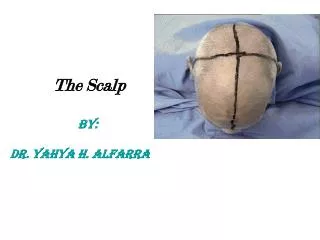
SCALP FLAPS & CRANIOTOMY PRINCIPLES
SCALP FLAPS & CRANIOTOMY PRINCIPLES. Scalp flaps- Historical perspective. Neolithic period in 2000 B.C 19 th century- trephines 1889 Wagner first osteoplastic bone flap Gigli saw for craniotomy- Obalinski in 1897 Electric and gas powered high speed drills.

SCALP FLAPS & CRANIOTOMY PRINCIPLES
E N D
Presentation Transcript
Scalp flaps-Historical perspective • Neolithic period in 2000 B.C • 19thcentury- trephines • 1889 Wagner first osteoplastic bone flap • Gigli saw for craniotomy- Obalinski in 1897 • Electric and gas powered high speed drills
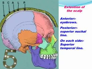
Anatomic and neurovascular considerations • 5 layers of scalp: Skin Connective tissue Aponeurosis Loose areolar tissue Pericranium
Land marks • Nasion • Bregma • Lambda • Inion • Pterion: Middle meningeal artery • Asterion: Transverse sigmoid junction
Fronto- temporal branch: anterior branch middle branch posterior branch • Middle division: 1 cm anterior to superficial temporal artery, subgaleal pad of fat Dissect between superficial and deep layers of superficial temporalis fascia
Blood supply • Superficial temporal artery • Occipital artery • Posterior auricular artery • Supra orbital and trochlear vessels
Principles of craniotomy • Preoperative review of patient • Preparation of scalp • Positioning of patient on the table • Scalp toilet • Marking of the incision • Draping
Planning • Position of lesion • Position of important structures • Contingency plan for enlarging incision • Obtain adequate closure
Principles • General principles: • Surgical exposure of the lesion • Neuro vascular supply • Cosmetic effect • Types: • Random pattern • Based on named vessel • Length not > 1.5 times base • Integrity of major vascular flap to be maintained • Incision in hair containing region • No crossed incisions
Principles • Skin incised with galea • Pressure over the scalp • Periosteum raised with scalp or separately • Raney’s clips, bipolar, Haemostatic artery forceps • Adequate retraction • Inner surface protected with moistened gauze • Roller gauze • Dissect in interfascial fat which is encountered within 4 cm of orbital rim
Types of craniotomies • Flap craniotomy • Trephine craniotomy • Flap craniotomy: • Osteoplastic • Free bone flap
Bone flaps • Most direct access to target • For cerebral convexity directly centered over the lesion • Number of burr holes varies • Separation of underlying dura • Beveling effect
Bone flaps • If dura is lacerated during cutting, saw should be turned off and removed backwards via entrance hole • Air cells opened: • Remove the mucosa • Pack with betadine soaked gelfoam • Pack with bone wax • Cover it up with vascularized tissue
Proposed bony cuts over venous sinuses should be done last- vascularity adherence • Cut sinus can be sewn/ tamponade • Bony bleeds stopped with bone wax • Penfield’s dissector to separate dura • Epidural tacking sutures to control epidural bleeding before opening dura • Others don’t in order to protect cortical blood vessels • Tailor to avoid dural venous channels
Opening of Dura mater • Manually palpate the dura • Dura opened as straight, curved or flap like incisions • Flaps based towards sinuses • Opened with sharp hook and knife • Incision further opened with dural scissors • Placement of cottonoid along the intended incision • Suitable cuff of dura around the bone for suturing later
Closure • Closure in layers • Check for BP- valsalva maneuver • Hitch suture • Water tight but not tension • Bone flap replacement • Skin closed in two layers
Bicoronal/ Souttarflap • Large exposures of anterior cranial fossa and sella • Fronto-temporal lesions and cranial base • Superior to zygomatic arch, 1 cm anterior to tragus- extends over the bregma to the corresponding site on the opposite side • Reflect up to orbit rim • Supraorbital/ trochlear vessels
Frontal/ Bifrontal bone flaps • Suitable for frontal lobe, sub-frontal approaches to anterior skull base, and trans cortical access to ventricles
Frontal/ Bifrontal bone flaps • An extended frontal or bi-frontal craniotomies for exposure of sella, anterior cranial base • Supine with head extended for these • Holes placed on either sides of sagittal sinus and intervening bone is removed with roneguers or drill • Either removed as single piece or conversion of frontal flap to bi-frontal flap • Combining a frontal flap with pterional flap
Frontal/ Bifrontal bone flaps • Goals of surgery dictate the craniotomy • Bilateral orbital craniotomies may be added to minimize frontal lobe retraction • Dural openings for a unilateral frontal craniotomy usually consist of flap reflected towards sagittal sinus • Superior sagittal sinus may have to be ligated
Frontal flap • Exposes anterior frontal lobe • Begins along coronal suture and curves anteriorly along the midline preferably ending at hair line
Temporal flap • Anterior temporal lobe and sub temporal access • Based on zygoma • Goes behind the ear • Extends anteriorly just behind the superior temporal line to the hair line
Link to video – • http://www.aiimsnets.org/NeurosurgeryAnimationVideoTemporalCraniotomy.html
Fronto- temporal (pterional) bone flap • Popularized by Yasargil • Most useful for aneurysms of anterior circulation, basilar top, also tumors of retro orbital, parasellar and subfrontal areas • Supine position with head end elevated to 30 degrees and rotated by the same to opposite side
Fronto- temporal (pterional) bone flap Link to video - http://www.aiimsnets.org/NeurosurgeryAnimationVideoPterionalCraniotomy.html
Fronto-temporal flap • Used for most pterional craniotomies • Combines frontal and temporal skin flaps • Extends from zygoma to 1-2 cm off the frontal midline following a curve behind the natural hair line • Temporalis muscle either dissected or reflected as a separate layer • In the later instance a cuff is left superiorly so as to suture it
Fronto- temporal (pterional) bone flap • Temporalis muscle dissected or reflected • Bone flap centered over the pterion • Key burr hole, frontal burr hole, posterior burr hole, last burr hole just above the zygoma • Further bone may be removed from the inferior temporal squama • To improve vision, drill the sphenoid ridge • Dural flap based on the orbit
FTOZ • Addition of orbito-zygomatic craniotomy will allow for a more lower and anterior approach • Suited for para-sellar, inter-peduncular lesions, • Basilar top aneurysm, • Carotico-ophthalmic aneurysms.
FTOZ Link to video - http://www.aiimsnets.org/NeurosurgeryAnimationVideoFrontotemporalOrbitozygomaticFTOZApproach.html
Question mark skin flap • Cranial trauma • Exposure to whole hemisphere • Based on zygoma • Blood supply from superior temporal and supra orbital vessels • Curves around 3.5 cm posterior to external auditory meatus • Anterior limb extends to hair line
Horse shoe skin flap • Expose any portion of cerebral convexity • Inverted “U” shaped with base directed towards vascular supply • Subtemporal exposure: anterior limb 1 cm anterior to the tragus • For anterior transcallosal approaches: over coronal suture
Mitre skin flap • Mitre hats worn by bishops • Occipital lobe, posterior falx and superior tentorial surface • Inion to vertex: vertical limb • Upper limb then falls over posterior parietal region towards the ear • Blood supply from the occipital artery
Linear and curvilinear incisions • Limited exposures • Simplicity • E.g.: MLSOC RMSOC Hockey stick incisions Linear incisions for temporal lobe & subtemporalaccess
CP angle tumors • Lateral • Prone • Three quarters prone • Sitting
Incision – Vertical linear ( 1 cm medial to the mastoid process ) ‘S’ / Lazy ‘S’ Inverted ‘J’ -shaped/ Hockey-stick Anatomical variants- Dolichoectatic VA/Occipital artery Hypoplastic VA (20 %)- Avoid extreme flexion Link to video - http://www.aiimsnets.org/NeurosurgeryAnimationVideoRetromastoidSuboccipitalCraniotomy.html Retromastoid suboccipital transmeatal approach
MLSOC Link to video - http://www.aiimsnets.org/NeurosurgeryAnimationVideoMidlineSuboccipitalCraniotomy.html
Poppens-Suboccipital Transtentorial Approach Link to video - http://www.aiimsnets.org/NeurosurgeryAnimationVideoPoppensSuboccipitalTranstentorialApproach.html