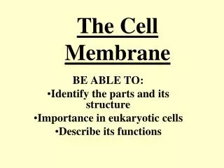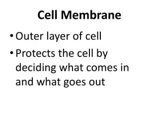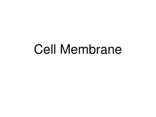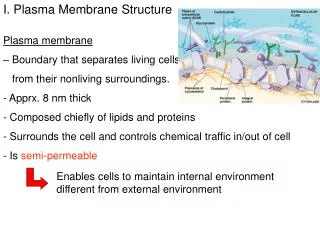
CELL MEMBRANE STRUCTURE & FUNCTIONS
CELL MEMBRANE STRUCTURE & FUNCTIONS. PASSIVE DIFFUSION, OSMOSIS, FACILITATED DIFFUSION AND ACTIVE TRANSPORT. WATER. Hydrophilic head. Hydrophobic tail. WATER. Phospholipid bilayer. Hydrophobic regions of protein. Hydrophilic regions of protein. TECHNIQUE. RESULTS. Extracellular

CELL MEMBRANE STRUCTURE & FUNCTIONS
E N D
Presentation Transcript
CELL MEMBRANE STRUCTURE & FUNCTIONS PASSIVE DIFFUSION, OSMOSIS, FACILITATED DIFFUSION AND ACTIVE TRANSPORT
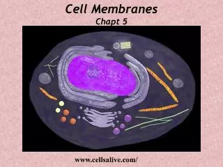
WATER Hydrophilic head Hydrophobic tail WATER
Phospholipid bilayer Hydrophobic regions of protein Hydrophilic regions of protein
TECHNIQUE RESULTS Extracellular layer Proteins Inside of extracellular layer Knife Cytoplasmic layer Plasma membrane Inside of cytoplasmic layer
Lateral movement (~107 times per second) Flip-flop (~ once per month) (a) Movement of phospholipids Fluid Viscous Unsaturated hydrocarbon tails with kinks Saturated hydro- carbon tails (b) Membrane fluidity Cholesterol (c) Cholesterol within the animal cell membrane
Cell Membrane Structures • Recognition Factors • Antenna that capture required solutes/hormnones • Amphipathic nature of the phospholipids and its consequences • Integral Proteins
Fibers of extracellular matrix (ECM) Carbohydrate Glyco- protein Glycolipid EXTRACELLULAR SIDE OF MEMBRANE Cholesterol Microfilaments of cytoskeleton Peripheral proteins Integral protein CYTOPLASMIC SIDE OF MEMBRANE
Signaling molecule Enzymes Receptor MEMBRANE PROTEINS ATP Signal transduction (b) Enzymatic activity (a) Transport (c) Signal transduction Glyco- protein (d) Cell-cell recognition (e) Intercellular joining (f) Attachment to the cytoskeleton and extracellular matrix (ECM)
EXTRACELLULAR SIDE N-terminus C-terminus CYTOPLASMIC SIDE Helix
ER 1 Transmembrane glycoproteins Secretory protein Glycolipid Importance1. Acquire molecules & Ions2.Transport into and out of cell through membrane3.Transport WITHIN the cell 2 Golgi apparatus Vesicle 3 Plasma membrane: Cytoplasmic face 4 Extracellular face Transmembrane glycoprotein Secreted protein Membrane glycolipid
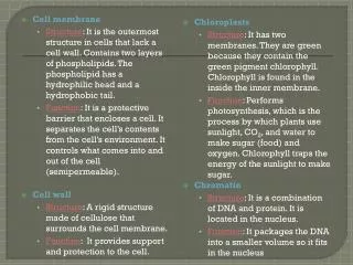
Cell Membrane Function Problems and Solutions 1. Relative concentrations a. Passive Transport b. Active Transport 2. Lipid bilayers are impermeable to most essential molecules and ions a. permeable to:
Cell membrane function, con’t b. impermeable to: - - -
DIFFUSION SIMPLE DIFFUSION REQUIRES A CONCENTRATION GRADIENT FACILITATED DIFFUSION REQUIRES A CONCENTRATION GRADIENT AND A PROTEIN TRANSPORTER
Molecules of dye Membrane (cross section) WATER Equilibrium Net diffusion Net diffusion (a) Diffusion of one solute
Relate to Lab: Glucose, Starch. NaCl, Proitein Equilibrium Net diffusion Net diffusion Net diffusion Net diffusion Equilibrium (b) Diffusion of two solutes
EXTRACELLULAR FLUID Channel protein Solute CYTOPLASM (a) A channel protein Solute Carrier protein (b) A carrier protein
ACTIVE TRANSPORT REQUIRES A PROTEIN TRANSPORTER AND ATP ENERGY
EXTRACELLULAR FLUID Na+ [Na+] high Na+ [K+] low Na+ Na+ Na+ Na+ Na+ Na+ ATP [Na+] low P Na+ P [K+] high CYTOPLASM ADP 2 3 1 K+ K+ K+ K+ K+ P K+ P 6 5 4
– EXTRACELLULAR FLUID + – ATP + H+ H+ Proton pump H+ – + H+ H+ H+ – + CYTOPLASM H+ – +
– + H+ ATP H+ – + H+ Proton pump H+ – + H+ H+ – + Diffusion of H+ H+ Sucrose-H+ cotransporter H+ – + Sucrose – + Sucrose
Passive transport Active transport REVIEW ATP Facilitated diffusion Diffusion
Osmosis • Passive Diffusion of Water Dissociation of ions in solution Aquaporins Osmotic concentration Free Energy Water Potential
Tonicity of cell IN COMPARISON TO the environment: The cell is ________in comparison to the environment. Hence, water will move from _____________________ to ____________. The results: Cell Interior: Less than 1% solute ENVIRONMENT 99% water
Hypotonic solution Isotonic solution Hypertonic solution H2O H2O H2O H2O (a) Animal cell Lysed Normal Shriveled H2O H2O H2O H2O (b) Plant cell Turgid (normal) Flaccid Plasmolyzed
Cell with less than 1 % SOLUTE (freshwater cell with more than 99% water) Cell with less than 1 % solute Environment with greater than 10% SOLUTE (less than 90 % water)
Environment: 0.01 M sucrose 0.01 M glucose 0.01 M fructose “Cell” 0.03 M sucrose 0.02 M glucose
Higher concentration of sugar Lower concentration of solute (sugar) Same concentration of sugar H2O Selectively permeable membrane Osmosis
Additional transport Mechanisms • Bulk Transport • Endocytosis • Phagocytosis • Pinocytosis • Receptor-mediated endocytosis
PHAGOCYTOSIS EXTRACELLULAR FLUID CYTOPLASM 1 µm Pseudopodium Pseudopodium of amoeba “Food” or other particle Bacterium Food vacuole Food vacuole An amoeba engulfing a bacterium via phagocytosis (TEM)
PINOCYTOSIS 0.5 µm Plasma membrane Pinocytosis vesicles forming (arrows) in a cell lining a small blood vessel (TEM) Vesicle
RECEPTOR-MEDIATED ENDOCYTOSIS Coat protein Receptor Coated vesicle Coated pit Ligand A coated pit and a coated vesicle formed during receptor- mediated endocytosis (TEMs) Coat protein Plasma membrane 0.25 µm