Download
1 / 45
450 likes | 507 Vues
Explore the latest in urolithiasis epidemiology, risk factors, diagnostic approaches, and medical management for better patient outcomes. Enhance your understanding with current guidelines and treatment decisions.

E N D
Medical management of urolithiasis 14 July 2015
Urolithiasis: iNTRODUCTION • Urolithiasis: A problem that has confronted clinicians since the time of Hippocrates & many family physicians have extensive experience in its clinical management • In recent years, technological advancements have greatly facilitated diagnosis of stone disease • Management of urolithiasis is also becoming increasingly well defined Portis AJ, et al. Am Fam Physician 2001;63:1329-38.
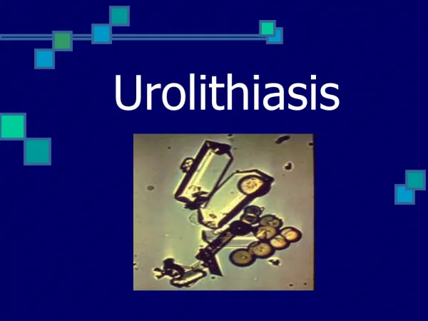
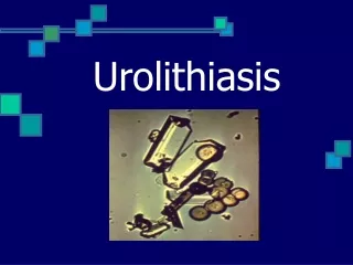
commonly occurring urinary tract stones and their salient features Kambadakone AR, et al. RadioGraphics 2010; 30:603–623.
Urolithiasis: Epidemiology The so-called stone belt (red) extends all the way around world & is characterized by urinary stone prevalence of 10 to 15% Fisang C, et al. Dtsch Arztebl Int 2015; 112: 83–91.
Urolithiasis: Epidemiology • 50 patients with previous urinary calculi: recurrence within 10 yrs • 2 to 3 times more common in males than in females • Occurs more often in adults than in elderly persons & more often in elderly persons than in children • Whites are affected more often than persons of Asian ethnicity, who are affected more often than blacks Portis AJ, et al. Am Fam Physician 2001;63:1329-38.
Urolithiasis: factors… • Occurs more frequently in hot, arid areas than in temperate regions • Decreased fluid intake & consequent urine concentration: most important factors influencing stone formation • Certain medications: triamterene, indinavir& acetazolamide • Dietary oxalate is another possible cause: • But role of dietary calcium is less clear & calcium restriction is no longer universally recommended Portis AJ, et al. Am Fam Physician 2001;63:1329-38.
Development of Urinary Calculi: RISK FACTORS Pietrow PK, et al. Am Fam Physician. 2006;74:86-94, 99-100.
Dependence of lithogenesis on urinary pH Fisang C, et al. Dtsch Arztebl Int 2015; 112: 83–91.
Relationship of Stone Location to Symptoms Portis AJ, et al. Am Fam Physician 2001;63:1329-38.
Plain film radiograph: calcium oxalate stone (arrow) in lower pole of rt kidney Pietrow PK, et al. Am Fam Physician. 2006;74:86-94, 99-100.
Radiograph: left kidney and upper bladder showing complete staghorn calculus Pietrow PK, et al. Am Fam Physician. 2006;74:86-94, 99-100.
Radiograph: a faintly radiopaque cystinecalculus (arrow) overlying 12th rib in left kidney of 13-yr-girl Pietrow PK, et al. Am Fam Physician. 2006;74:86-94, 99-100.
Initial management of radiologically confirmed urolithiasis (KUB = kidney, ureters and bladder)
Medical Management of Nephrolithiasis Pietrow PK, et al. Am Fam Physician. 2006;74:86-94, 99-100.
Algorithm: management of urolithiasis during pregnancy Masselli G, et al. Insights Imaging. 2014; 5: 691–696.
Probability of Stone Passage* Portis AJ, et al. Am Fam Physician 2001;63:1329-38.
TREATMENT DECISION BASED ON STONE LOCATION: KIDNEY Kambadakone AR, et al. RadioGraphics 2010; 30:603–623.
TREATMENT DECISION BASED ON STONE LOCATION: URETER Kambadakone AR, et al. RadioGraphics 2010; 30:603–623.
TREATMENT DECISION BASED ON STONE COMPOSITION Kambadakone AR, et al. RadioGraphics 2010; 30:603–623.
PROPHYLACTIC MANAGEMENT IN URINARY STONE FORMER • All patients presenting with renal colic should be given general medical advice to decrease risk of future stone episode • This advice includes increasing fluid intake so that urine output is > 2L/day • Best estimation of urine output is clear looking urine. This prevents urine stagnation & thus decreases stone risk Spernat D, et al. BJU International. 2011; 108: 9-13.
PROPHYLACTIC MANAGEMENT IN URINARY STONE FORMER • Patients should their urinary citrate level through consumption of citric juices • Urinary citrate: A potent stone inhibitor • Citrate binds calcium in urine, decreasing supersaturation& growth of crystals • in urinary citrate level even in patients with normal urinary citrate helps prevent stone recurrence • Purine intake (animal meat) should be moderated as it urinary calcium, oxalate & uric acid secretion • Restricting animal protein & salt while maintaining normal calcium intake stone recurrence rates compared with low calcium diet Spernat D, et al. BJU International. 2011; 108: 9-13.
PROPHYLACTIC MANAGEMENT IN URINARY STONE FORMER • High sodium load can risk of calcium oxalate stone formation & thus salt restriction is also advised • Obesity risk of stone disease by urinary acidity, hypocitraturia& hyperuricosuria • Weight loss & low fat diet should be encouraged. This is especially important in patients with bowel disease or malabsorptiveconditions Spernat D, et al. BJU International. 2011; 108: 9-13.
PROPHYLACTIC MANAGEMENT IN URINARY STONE FORMER • Calcium intake within dietary recommendations should be continued even in patients with calcium oxalate stones • Low dietary calcium leads to increased unbound oxalate to be reabsorbed in the gut & thus further risk of calcium oxalate stones • Patients should also avoid excessive (maximum daily dose 2 g) vitamin C supplements, as this can increase oxalate excretion Spernat D, et al. BJU International. 2011; 108: 9-13.
PROPHYLACTIC MEDICATIONS: Thiazide diuretics • Thiazides stimulate calcium reabsorption in distal nephron while promoting excretion of sodium. This urinary calcium excretion but may lead to hypokalemia • Hypokalemiacan in turn cause hypocitraturia. Thus potassium citrate supplementation (40 – 60 mEq/day) is recommended with thiazide diuretics • A recent Cochrane review has demonstrated that, in patients with idiopathic hypercalciuria & recurrent stones: Addition of thiazides to normal or modified diet stone recurrence & formation rate Spernat D, et al. BJU International. 2011; 108: 9-13.
PROPHYLACTIC MEDICATIONS: Potassium citrate • Potassium citrate has several features • It maintains urine pH above pKa for uric acid thus promoting dissolution of uric acid crystals. This uric acid & calcium stone formation by formation of a nidus • Citrate also directly prevents complexation of calcium • In patients with either hypocitraturiaor acidic urine pH, treatment with this medication urinary citrate levels, pH and potassium. This is associated with remission rate of stone disease of up to 91% Spernat D, et al. BJU International. 2011; 108: 9-13.
PROPHYLACTIC MEDICATIONS: Allopurinol • Allopurinol inhibits xanthine oxidase converting xanthine to uric acid& therefore uric acid production & hyperuricosuria • This in turn spontaneous nucleation of calcium oxalate • Randomized controlled trials have demonstrated that allopurinol stone recurrence in patients with idiopathic calcium oxalate stones who had hyperuricosuria • Allopurinol is therefore effective in pure uric acid calculi & calcium based calculi Spernat D, et al. BJU International. 2011; 108: 9-13.
PROPHYLACTIC MEDICATIONS: UroPhos-K • UroPhos-K: A slow release neutral form of potassium phosphate • It produces sustained hypocalciuric response & maintains bone mass in patients with absorptive hypercalciuria • This effect is achieved by directly impairing renal tubular reabsorption of calcium & by binding calcium in gut • It also raises urine pH • UroPhos-K causes a sustained & marked in urinary calcium. This effect occurs by combination of intestinal absorption, bone reabsorption & improved renal calcium reabsorption • Well tolerated compared with placebo Spernat D, et al. BJU International. 2011; 108: 9-13.
PROPHYLACTIC MEDICATIONS: Sodium bicarbonate • Major goal of therapy: To urine pH above 5.5 & preferably to 6.5 – 7.0. This treatment enhances dissociation of uric acid & inhibits uric acid stone formation • However, treatment may be complicated by calcium oxalate (due to sodium load) or calcium phosphate (due to pH above 7.0) stone formation • Thus potassium citrate is preferable as it avoids sodium load that may precipitate calcium oxalate stone formation Spernat D, et al. BJU International. 2011; 108: 9-13.
DISSOLUTION THERAPY • Oral Medications: • Sodium bicarbonate • Potassium citrate • Percutaneous Instillation: • Calcium oxalate stones are resistant to dissolution therapy. However, struvitecalculi have been associated with (limited) successful dissolution therapies since 1943 (Suby’ssolution G). The following two solutions are still used in limited cases: • Hemiacidrin/renacidrin • Tham E Spernat D, et al. BJU International. 2011; 108: 9-13.
MEDICAL EXPULSION THERAPY • Medical expulsion therapy (MET): Beneficial for distal ureteric calculi • No evidence that MET improves spontaneous stone passage rate of proximal ureteric calculi • However, tamsulosin has been shown to significantly passage of stones between 5 & 10 mm from proximal to distal ureter Spernat D, et al. BJU International. 2011; 108: 9-13.
High Fluid intake • A large diuresis that pushes stone into bladder sounds plausible; however, studies have demonstrated that it is actually more likely to counteract passage of stone & to cause more pain • Cochrane database review from 2005: No evidence to support diuresis as method of pain relief or stone expulsion Spernat D, et al. BJU International. 2011; 108: 9-13.
Alpha blockers • Alpha blockers inhibit ureteral muscle contraction, reduce basal tone& decrease peristaltic frequency and colic pain facilitating ureteral stone expulsion • Meta-analysis has demonstrated: Advantage to alpha blocker use that it increases spontaneous stone passage rate by 14 – 29% which is statistically significant. This is the recommended MET (combined AUA and European Association of Urology (EAU) guidelines 2007) Spernat D, et al. BJU International. 2011; 108: 9-13.
Alpha blockers: Spontaneous stone passage at one month Tamsulosin is most commonly used and studied agent Spernat D, et al. BJU International. 2011; 108: 9-13.
Use of alpha-blockers in patients with ureteral stones results in higher stone-free rate & shorter time to stone expulsion Alpha-blockers should therefore be offered as part of medical expulsive therapy as 1 of primary treatment modalities
Principal substances used in medicinal prophylaxis of urinary stones Fisang C, et al. Dtsch Arztebl Int 2015; 112: 83–91.
Principal substances used in medicinal prophylaxis of urinary stones Fisang C, et al. Dtsch Arztebl Int 2015; 112: 83–91.
Principal substances used in medicinal prophylaxis of urinary stones Fisang C, et al. Dtsch Arztebl Int 2015; 112: 83–91.
Salient Features of Various Urologic Interventional Procedures for Urolithiasis Kambadakone AR, et al. RadioGraphics 2010; 30:603–623.
Salient Features of Various Urologic Interventional Procedures for Urolithiasis Kambadakone AR, et al. RadioGraphics 2010; 30:603–623.
Salient Features of Various Urologic Interventional Procedures for Urolithiasis Kambadakone AR, et al. RadioGraphics 2010; 30:603–623.
COMPLICATIONS OF UROLITHIASIS • Renal failure • Ureteral stricture • Infection, sepsis • Urine extravasation • Perinephric abscess • Xanthogranulomatous pyelonephritis Portis AJ, et al. Am Fam Physician 2001;63:1329-38.