
Analysis of M-phase Nuclei in Non-Ventricular and Ventricular Tissue Using p-Histone Staining
This study investigates the distribution and characteristics of M-phase nuclei in both non-ventricular and ventricular tissues, employing p-histone staining techniques. Using various control and experimental models, we analyze the percentage of M-phase nuclei to assess cellular proliferation in these regions at different developmental stages (E13.5 and E14.5). We present findings that illustrate significant variations in M-phase activity, contributing to our understanding of cell cycle regulation in cardiac development.
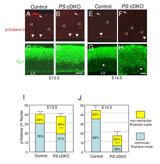

Analysis of M-phase Nuclei in Non-Ventricular and Ventricular Tissue Using p-Histone Staining
E N D
Presentation Transcript
20% 24% 49% *** 76% 80% 51% 62% *** PS cDKO Control PS cDKO Control A B E F p-histone 3 G H C D Tuj-1 LV LV E14.5 E13.5 I J 50 50 E13.5 E14.5 40 40 non-ventricular M-phase nuclei 30 30 p-histone 3+ Nuclei 20 20 ventricular M-phase nuclei 10 10 38% 0 0 Control PS cDKO Control PS cDKO