Cardiovascular Development
Cardiovascular Development. Matt Velkey jvelkey@umich.edu 454A Davison Reading: Langman’s Medical Embryology, 11 th ed. Ch 12 pp. 165-200. The cardiovascular system is mesodermally derived. Specifically, lateral splanchnic mesoderm…. Gilbert fig 14.1.

Cardiovascular Development
E N D
Presentation Transcript
Cardiovascular Development Matt Velkey jvelkey@umich.edu 454A Davison Reading: Langman’s Medical Embryology, 11th ed. Ch 12 pp. 165-200
The cardiovascular system is mesodermally derived Specifically, lateral splanchnic mesoderm… Gilbert fig 14.1
angiogenic cell clusters (angioblasts/hemangioblasts) (right endocardial tube) (right dorsal aorta) pericardial cavity cardiogenic field The cardiogenic field is established in the mesoderm just after gastrulation (~18-19 days) and develops into a fully functional, multi-chambered heart by the 8th week blood islands (developing blood vessels) Langman’s fig 12-1
Carlson fig 6-14 Establishment of the heart fields BMP2 & 4 in mesoderm WNT inhibition (from “Anterior” Visceral Endoderm) in anterior mesoderm BMP2,4+/Wnt- expression pattern specifies cardiac tissue (evidenced by expression of NKX-2.5, aka tinman) Langman’s fig 12-9
Repositioning the cardiogenic field (mammals) Larsen’s fig 12-6 Langman’s fig 12-6
Cranial folding Cranial folding rotates cardiogenic area, moves it ventrally and caudally, and orients heart tube and pericardial cavity Moore & Persaud fig 13-9
Lateral folding Moore & Persaud fig 13-8 Lateral folding apposes paired heart tube primordia and brings dorsal aortae to midline Heart primordia fuse to form tubular heart
Fusing cardiac primordia “conotruncus” (outflow tract) future ventricles future atria 22 days 21 days Langman’s fig 12-7 septum transversum (liver & diaphragm primordium)
Retinoic acid (RA) and other factors determine the cranio-caudal axis of heart primordia • Primary heart field: left ventricle • Posterior segment of primary heart field: atria • Secondary heart field: right ventricle & outflow tract • EXOGENOUS RETINOIDS CAN INTERFERE WITH THIS PROCESS! Carlson fig 17-17
Heart tube and dorsal aortae in place Transverse pericardial sinus Langman’s fig 12-5
22 days 30 days …to this? Heart folding How do you get from this… Carlson fig 17-19 Langman’s fig 12-7
aortic roots truncus arteriosus bulbus cordis ventricle atrium sinus venosus Folding and rotation of heart tube • Ventricle moves ventrally and to right • Atrium moves dorsally and to left 22 days 23 days 24 days Langman’s fig 12-6
Folding and rotation of heart tube QuickTime version • Ventricle moves ventrally and to right • Atrium moves dorsally and to left
Looped heart anatomy Carlson fig 17-18
At 30 days… • Atrial partitioning just beginning to happen • Primary interventricular foramen undivided • Outflow tract (bulbus cordis/truncus arteriosus) undivided • Proximal portion of bulbus cordis becomes trabeculated and forms the right ventricle • Atrioventricular canal undivided Langman’s fig 12-8
Partitioning the AV canal Atrioventricular Endocardial Cushions • Retinoic acid (and many other factors) dependent* conversion of endocardial endothelium in the AV canal into mesenchyme that proliferates to form “cushions” of tissue • Dorsal and ventral cushions fuse in the middle to form a block of tissue that divides right and left AV canals Moore & Persaud fig 13-11 *can be disrupted by exogenous retinoids and/or in many genetic disturbances (e.g. trisomy 21 –aka Down Syndrome)
Partitioning the AV canal based on Larsen’s fig 12-27 QuickTime version • Atrioventricular Endocardial Cushions • Expansion of tissue around periphery of AV canal • Dorsal and ventral cushions fuse to form right and left AV canals divided by septum intermedium (failure of cushions to fuse causes “persistent AV canal”) • Remodeling brings right and left AV canals into alignment with future right and left ventricles –movement driven by differential growth (failure of this process leads to “double inlet” defects)
Persistent A-V canal • Failure of A-V endocardial cushions to fuse • Common in Down syndrome View of A-V canal looking down into ventricles with atria removed (e.g. at dotted line on left) Carlson fig 17-32 Fig 174, Nishimura & Okamoto (1976)
Atrial septum formation 33 40 43 • septum primum • ostium primum • ostium secundum • septum secundum • foramen ovale Larsen’s figs 12-24, 12-25, 12-26
septum primum • ostium primum • ostium secundum • septum secundum • foramen ovale QuickTime version from Larsen’s figs 12-24, 12-25, 12-26 Atrial septum formation
Why is a right-left shunt necessary? In the fetus, blood is oxygenated in the placenta and delivered to the heart via the inferior vena cava: • Shunted to left atrium via foramen ovale • Also shunted from pulmonary outflow via ductus arteriorsus Carlson fig 17-19
Atrial Septal Defects (ASDs) Multi-factoral, so many etiologies, but one well-known example is mutation in NKX2-5 characterized by: • Incomplete septum secundum • AV “bundle block” (disruption of conducting fibers, evidenced by prolonged P-R interval on ECG) –typically worsens with age Schott et al. (1998). Science. 281:108-111 Doppler flow echo-CG showing RA to LA shunt ECG showing progressive AV block (normal P-R int. 0.13-0.2s)
Late 5th week • Atrium in process of septal formation • Interventricular septum still incomplete • muscular septum present • membranous septum ABSENT • Outflow tract still undivided Moore & Persaud fig 13-17 membranous septum will form here muscular septum Langman’s fig 12-17
Outflow Tract Partitioning Membranous septum formed by contributions from AV cushions and truncoconal cushions. • Truncoconal ridges • Neural crest-derived endocardial cushions form in truncus arteriosis and conus (bulbus) cordis region • Fuse at truncoconal transition and “zip” proximally and distally to form aorticopulmonary septum. R L 5th wk 7th wk 9th wk QuickTime version 8th wk Larsen’s fig 12-33
Outflow Tract Defects • Typically due to failure of neural crest-derived conotruncal cushions • Associated with other disorders affecting neural crest: DiGeorge syndrome, fetal alcohol syndrome, chromosome 22 mutations (e.g. Tbx-1) • Can also arise due to defects in secondary heart field (e.g. Hand-2, retinoids) • Examples include: persistent truncus arteriosus, transposition of the great vessels, aortic and/or pulmonary stenosis (tetrology of Fallot)
Persistent Truncus Arteriosus (left)Great Vessel Transposition (right) Carlson fig 17-31
Pulmonary Stenosis: Tetrology of Fallot • Pulmonary stenosis • Overriding aorta • Intraventricular septal defect • Hypertrophy of right ventricle • (Patent ductus arteriosus)… so really a “pentology” Carlson fig 17-40
Other signs of Tetrology of Fallot • Respiratory distress (dyspnea), with rapid breathing • Characteristic squatting posture, at rest, to relieve dyspnea • Coeur en sabot(boot-shaped heart) in Xray • Cyanosis develops gradually, with intermittent severe “spells” • Clubbed fingers and toes with cyanosis
Formation of semilunar valves also outflow cushion dependent… P L Pulmonary valve tubercles R Aorta A Pulmonary artery Truncoconal septum Larsen’s fig 12-34 Tricuspidvalve Langman’s fig 12-27 Mitral valve Langman’s fig 12-23
Formation of AV valves dependent on AV cushions and ventricular myocardium… Moore & Persaud fig 13-19
Mitral stenosis/hypoplastic left ventricle • Failure of left A-V valve (tricuspid valve) to form: LV and aorta becomes hypoplastic because of reduced load. • OK in embryo since oxygenated blood is coming from IVC and can be distributed systemically via ductus arteriosus. • But, this arrangement doesn’t work so well in a breathing infant since oxygenation occurs in the lungs.
Fontan Repair of Single Ventricle • Reroute systemic venous blood to lungs for oxygenation • Reroute right ventricle outflow to aorta • Oxygenated blood from lungs flows from LA in RA via patent foramen ovale and RV pumps oxygenated blood out to body.
Blood Vessel Development Langman’s fig 16-04
1 2 3 4 6 Aortic arches at pharyngeal arches and pouches Each pharyngeal arch has aortic arch, cranial nerve, and cartilage components… 7 weeks 4th arch 6th arch 1st arch 2nd arch • Five aortic arches are forming during the 4th and 5th weeks. • 5th arch fails to form; arches are numbered: 1, 2, 3, 4, and 6 3rd arch “branchial” = pharyngeal Langman’s fig 16-04
6 months postnatal 6 weeks From this… …to this Changes in the aortic arch pattern Animation: http://www.indiana.edu/~anat550/cvanim/aarch/aarch.html Moore & Persaud fig 13-39
Changes in the aortic arch pattern (AS,1,2,3,5) Animation: http://www.indiana.edu/~anat550/cvanim/aarch/aarch.html • Aortic Sac (AS): • proximal part of aortic arch • brachiocephalic a. • 1st arch mostly disappears • maxillary a. • (part of external carotid a.?) Aortic sac • 2nd arch mostly disappears • stapedial a. • (hyoid a.?) • 3rd arch: • common carotid a. • part of internal carotid a. • internal and external carotid aa. sprout from 3rd arch Moore & Persaud fig 13-39 5th arch fails to form
Changes in the aortic arch pattern (4) Animation: http://www.indiana.edu/~anat550/cvanim/aarch/aarch.html • 4th arch on right: • proximal segment of right subclavian a. (rest of subclavian a. from 7th intersegmental a. and R dorsal aorta) • 4th arch on left: • arch of aorta (from left common carotid a. to left subclavian a. only) Moore & Persaud fig 13-39
Changes in the aortic arch pattern (6) Animation: http://www.indiana.edu/~anat550/cvanim/aarch/aarch.html (6th arch = pulmonary arch) • 6th arch on left: • left pulmonary artery • distal segment persists as ductus arteriosus • 6th arch on right: • right pulmonary artery • distal segment regresses Moore & Persaud fig 13-39
Aortic Arch Anomalies normally regresses • Double aortic arch abnormal persistence of right distal segment ~1:1000 incidence –often assoc. with dysphagia and/or dyspnea • Right aortic arch abnormal persistence of right distal segment & regression of left distal segment ~1:1000 incidence –usually asymptomatic • Aberrant right subclavian (from aortic arch) (abnormal regression of rightproximal segment & persistence of rightdistal segment) ~1:100 incidence –often assoc. with dysphagia and/or dyspnea; also, R radial pulse may be weak normally persists normally persists Carlson fig 17-42
Interrupted aortic arch (IAA) • Abnormal regression of proximal left 4th arch • Output to left upper limb, trunk, and both lower limbs is via pulmonary trunk (connected to descending aorta via ductusarteriosus) • Rather asymptomatic at first, but ductus starts to close during first 2 weeks of life, so needs to be caught and fixed (via surgical reconstruction) by then • Neural crest etiology: Rare (1:50,000) in general population, but rather common (~10%) in patients with DiGeorge syndrome (22q deletion) R and L common carotid arteries and right limb* R subclavian a. *The R subclavian is shown here as a separate branch from the arch of the aorta; the middle vessel is the R common cartotid; the remaining vessel coming from the arch of the aorta is the L common carotid. L subclavian a. Carlson fig 17-44
Coarctation of aorta • Collateral circulations can compensate for postductal coarctation • But, not perfect, so blood pressure in upper limbs is higher compared to lower limbs • Preductal coarctation is MUCH less common (5% of coarctations) ~1:3000 incidence overall, but commonly coincident w/ Turner’s syndrome (~20%) and neural crest disorders Moore & Persaud, Fig 15-29; ref to Sadler, Fig 11-37
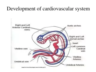
Venous system development cardinal veins sinus venosus vitelline veins umbilical veins Langman’s fig 12-41
Vitelline and umbilical veins change during liver development sinus venosus cardinal V L hepatocardiac channel hepatic sinusoids L vitelline V duodenum umbilical V hepatic portion of inferior vena cava hepatic V(R vitelline V) 4 weeks duodenum hepatic V 5 weeks yolk sac Langman’s fig 12-42 R hepatocardiac channel ductus venosus • R hepatocardiac channel hepatic portion of IVC • R umbilical V regresses • proximal L umbilical V regresses • distal L umbilical V persists and then round ligament of the liver (ligamentum teres hepatis) • ductus venosus ligamentum venosum portal V superior mesenteric V 6 weeks 8 weeks splenic V Langman’s fig 12-43
Systemic venous development posterior cardinal veins anterior cardinal veins Langman’s fig 12-41
5.5 weeks Systemic venous development: shift to the right 6 weeks 5 weeks Moore & Persaud
L. brachiocephalic anastomosis 7 weeks Adult Systemic venous development: shift to the right Moore & Persaud What if the brachiocephalic anastomosis fails?
Double superior vena cava Left superior vena cava Langman’s fig 12-46 viewed from behind
Absent inferior vena cava Double inferior vena cava Langman’s fig 12-45