Download
1 / 1
10 likes | 180 Vues
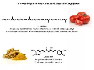
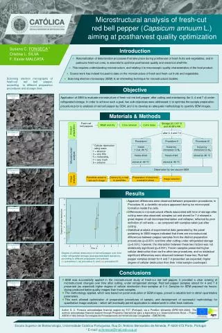
Procedure I 0 d. Procedure I 7 d. Procedure II 0 d. Procedure II 7 d. Procedure III 0 d. Procedure III 7 d. Microstructural analysis of fresh-cut red bell pepper ( Capsicum annuum L.) aiming at postharvest quality optimization. Susana C. FONSECA * Cristina L. SILVA

E N D
Procedure I 0 d Procedure I 7 d Procedure II 0 d Procedure II 7 d Procedure III 0 d Procedure III 7 d Microstructural analysis of fresh-cut red bell pepper (Capsicum annuum L.) aiming at postharvest quality optimization Susana C.FONSECA * Cristina L. SILVA F. Xavier MALCATA Introduction • Rationalization of deterioration processes that take place during postharvest of fresh fruits and vegetables, and in particular fresh-cut ones, is essential to optimize postharvest quality and maximize shelf-life. • This requires understanding microstructure, and relating it to macroscopic quality characteristics of the food product. • Scarce work has indeed focused to date on the microstructure of fresh and fresh-cut fruits and vegetables. • Scanning electron microscopy (SEM) is an interesting technique for microstructural studies. Scanning electron micrographs of fresh-cut red bell pepper, according to different preparation procedures and storage time: Objective Application of SEM to evaluate microstructure of fresh-cut red bell pepper, after cutting and maintaining (for 0, 4 and 7 d) under refrigerated storage. In order to achieve such a goal, two sub-objectives were addressed: i) to optimise the sample preparation procedure prior to analysis of red bell pepper by SEM, and ii) to develop an adequate methodology to quantify SEM images. Materials & Methods Fresh red bell peppers Wash and dry Core removal Cut in dices Storage (2 ± 0.5 C and 90-95% RH Product preparation after 0, 4 and 7 d Procedure I Procedure II Procedure III * Cellular destruction rating scale: 1 = absence 3 = very little 5 = moderately, 7 = very much 9 = extreme frozen 1 d at -80 °C frozen by immersion in N2 frozen by immersion in N2 Microstructural analysis freeze-dried freeze-dried stored at -80 °C stored at -80 °C stored at -80 °C Observation by low vacuum SEM Image analysis Delivery by e-mail to panellists Preparation of electronic evaluation sheet Image selection Panellists asked to rate each image* Results • Apparent differences were observed between preparation procedures; in Procedure III, a dendritic structure appeared during ice microcrystal formation inside the cells. • Differences in microstructural effects associated with time of storage after cutting were also observed; samples cut and stored for 7 d showed a great degree of cell decompartmentation and collapse, reflected by poor definition of cell walls — as compared with samples taken just after cutting. • Statistical analysis of experimental data generated by the panel pertaining to SEM images indicated that there are microstructural differences between pepper samples from the distinct preparation procedures (p=0.001) and time after cutting under refrigerated storage (p=0.001); however, the interaction between these two factors was not statistically significant (p=0.051). Frozen samples presented higher cellular destruction than via the other two procedures, and no statistical significant differences were observed between these two. Red bell pepper samples stored for 4 and 7 d presented (as expected) higher degree of cellular destruction than their initial sample counterpart. 9 8 7 6 Degree of cellular destruction 5 4 3 2 1 0 1 2 3 4 5 6 7 Time, d Degree of cellular destruction of red bell pepper over time under refrigerated storage (average±standard deviation), according to different preparation procedures: (●) procedure I; (■) procedure II; and (▲) procedure III. Conclusions • SEM was successfully applied in the microstructural study of fresh-cut red bell pepper; it provided a clear viewing of microstructural changes over time after cutting, under refrigerated storage. Red bell pepper samples stored for 4 and 7 d presented (as expected) higher degree of cellular destruction than samples at 0 d. Samples for SEM prepared via freeze-drying produced better quality images than frozen samples. • The methodology applied, which was based on panel evaluation of SEM images, proved a valuable tool to obtain quantitative parameters. • This work allowed optimization of preparation procedures of sample, and development of successful methodology for quantitative image analysis – which will eventually permit application to related work in other food matrices. Author S. C. Fonseca acknowledges financial support by FCT (Portugal), via a PostDoc fellowship (BPD/1601/2000). The authors acknowledge financial support through Programa Operacional para a Agricultura e o Desenvolvimento Rural – Project AGRO nº 822 (Novas Tecnologias de Processamento de Hortofrutícolas Congelados – EMERCON). Escola Superior de Biotecnologia, Universidade Católica Portuguesa, Rua Dr. António Bernardino de Almeida, P-4200-072 Porto, Portugal * E-mail: scfonseca@mail.esb.ucp.pt