Download
1 / 24
250 likes | 539 Vues
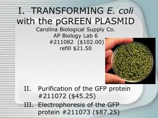
I. TRANSFORMING E. coli with the pGREEN PLASMID Carolina Biological Supply Co. AP Biology Lab 6 #211082 ($102.00) refill $21.50. Purification of the GFP protein #211072 ($45.25) Electrophoresis of the GFP protein #211073 ($87.25). Purpose.

E N D
I. TRANSFORMING E. coli with the pGREEN PLASMIDCarolina Biological Supply Co.AP Biology Lab 6 #211082 ($102.00)refill $21.50 Purification of the GFP protein #211072 ($45.25) Electrophoresis of the GFP protein #211073 ($87.25)
Purpose Part 1 - To transform E. coli (plasmid contains ampicillin resistance and green fluorescent protein from Aequorea victoria (a bioluminescent sea jelly)) Part 2 - To purify the GFP protein produced by the newly transformed E. coli Part 3 - To use electrophoresis to verify the presence of the purified GFP protein.
Teacher Prep-Part I • Pour LB agar plates, or prepare lab to have students pour plates. • Inoculate starter plates 24 hrs prior to the lab • Set up student stations. • Set water bath to 42°C.
Background Information • An understanding of the characteristics and requirements of bacteria including the effects of antibiotics. • An understanding of both natural and artificial transformation in bacteria. • An understanding of genetic engineering including the controversy. Excellent opportunity to discuss/ debate bioethics. • Techniques: Use of sterile techniques (TREAT ALL BACTERIA AS PATHOGENS), burel pipet, little finger technique, micropipet, and experience loading electrophoresis gels. • Complete prelab: Make predictions about outcome.
Procedure - Part ITransformation • Mark the 2 transformation tubes:+ & - • Add 250 uL CaCl2 (ice cold) to each tube. • Using a sterile inoculating loop, add E. coli to each tube. MIX WELL!!! • Using a new inoculating loop, add 1 loop of plasmid to + tube. • Incubate both tubes on ice for 15 minutes.
Procedure - Part ITransformation • Label media:
Procedure - Part ITransformation • Heat shock the cells for 90 sec. Tubes should go directly from ice to heat, and then directly back to ice. • Using the same burel pipet feed the cells in each tube with 250 uL of Luria broth. (Best to feed - tube first) • Room/ body temperature 5 - 15 minute recovery period. • Using a new pipet transfer 100 uL of - cells to each of the - plates. • Using a new pipet transfer 100 uL of + cells to each of the + plates. • Spread the bacteria using the glass beads. • Clean up.
Results • Plates without ampicillin (#1 & 2) will show a “lawn” of growth: All materials needed for growth are present without poison, so bacteria will grow. • Ampicillin plate without plasmid (#3): Bacteria is killed - no growth. • Ampicillin plate with plsamid (#4) will show individual green (transformed) colonies that glow under UV light: Only bacteria that was transformed will grow; therefore, the lesser growth will allow individual colonies to be seen.
II. Purification of the GFP protein #211072 ($45.25) • Purpose: To purify the GFP protein produced by the newly transformed E. coli
Teacher Prep1 day before lab • Add ampicillin to Luria broth • Prepare overnight cultures of GFP expressing E. coli colonies (Taken from Plate 4 of part 1 no more than 2 - 3 days old) by inoculating 2 mL portions of Luria broth w/ amp. Incubate at 33C (37C). • Equilibrate the HIC bead resin. • Set up student stations.
Supplies • In Kit • Needed
Background Information • Using micropipets
Procedure - Part IIPurification of GFP by HIC • Collect cells in pellet
Procedure - Part IIPurification of GFP by HIC • Collect GFP Extract from Cell Pellet
Procedure - Part IIPurification of GFP by HIC • Collect GFP Extractfrom Cell Pellet
III. Electrophoresis of the GFP protein #211073 ($87.25)Purpose: Part 3 - To use electrophoresis toverify the presence of the purified GFP protein.
Teacher Prep • Prepare tris-glycine SDS buffer • Prepare a boiling beaker of water or a water bath or heating block at 95C • Aliquot protein marker in 7 uL portions (1/ gel) • Set up student workstation
Student Workstations • 2 clean 1.5-mL microcentrifuge tubes • students’ tube of purified GFP protein (on ice) • students’ tube of cell lysate (on ice) • pipet and pipet tips (2–20 μL) • permanent marker
The following materials will be shared by every two or three groups: • 1 tube of protein markers (on ice) • 12% polyacrylamide gel • protein gel electrophoresis apparatus and power supply • 1x tris-glycine-SDS running buffer • staining tray • tube of 4ー— loading dye • spatula for separating gel plates • gloves • Students will share the following: • equipment for maintaining 95ーC • COOMASSIEョ blue protein staining solution • white light transilluminator (optional) • instant or digital camera (optional)
Procedure I. Denature Proteins, Load Gel, and Electrophorese (1 hour, 30 minutes) • Note: There are 10 wells in each gel and each group will load two samples, so each gel will be shared by two or three groups. The protein markers also will be shared. • 1. Place the 12% polyacrylamide gel into an appropriate gel electrophoresis apparatus and add 1 x tris-glycine-SDS buffer to both chambers. Remove the comb slowly and carefully. Make sure that the sides of the wells are straight. • 2. Use a permanent marker to label two 1.5-mL tubes: • CL = cell lysate (from Part A, Step 9) • GFP = purified GFP (from Part A, Step 16)
3. Transfer 5 μL of cell lysate (CL) and 15 μL of purified GFP (GFP) into the appropriate tubes. These volumes correspond to approximately 10 μg of total protein. Make sure that you have a tube containing protein markers (PM). • 4. Add 1.6 μL of 4ー— loading dye to tube CL. Add 5 μL of 4ー— protein loading dye to tube GFP. • 5. Heat the samples for two minutes at 95ーC to denature the proteins. Do not heat the marker. The marker sent with this kit is formulated to be loaded without being heated. • 6. Refer to Figure 1. Load 5μL of the protein marker into the second well. • Load the entire contents of each sample tube into a separate well in the gel. Use a fresh tip to load each sample. Load your samples in the following order from left to right: PM (5 μL), CL (group 1), GFP (group 1), CL (group 2), GFP (group 2), CL (group 3), GFP (group 3). Do not use the first and last lanes of the gel, because bands from samples run in these lanes can become distorted.
7. Close the tank of the protein gel electrophoresis unit. Connect themelectrical leads to a power supply, anode to anode and cathode to cathode. • Electrophorese at 175 volts until the bromophenol blue band has moved to the bottom of the gel. This should take approximately one hour. • 8. Turn off the power supply, disconnect the leads from the inputs, and remove the top of the electrophoresis box. • 9. Carefully remove the gel cassette from the electrophoresis chamber. Open the cassette with a spatula and transfer the gel to a staining tray.Wear gloves when you handle the gel. Handle the gel with care to avoid tearing it.
II. Stain the Gel with COOMASSIEョ Blue, View, and • Photograph (3+ hours) • 1. Flood the gel with COOMASSIEョ blue protein staining solution, cover the staining tray to prevent evaporation, and allow the gel to stain from one hour to overnight. • 2. Wearing gloves, pour off the staining solution. Remove the excess stain by rinsing the gel in tap water. • 3. Destain the gel in tap water for one to two hours, changing the water every 15 minutes. (The gel may also be destained overnight in a small volume of water.) Protein bands will appear blue. The longer the destaining period, the less intense the background blue stain will appear. Do not destain for too long or the protein bands may lose their intensity. • 4. View the gel on a white background or on a white light transilluminator. • 5. Photograph the gel with an instant or digital camera (optional).