
Double Fluorescence Staining Reveals Subpopulation of Large Neurons in Neurofilament Analysis
This study presents double fluorescence staining for neurofilament (RT97, blue) and α3 (red), highlighting a distinctive subpopulation of large neurons. The α3 staining outlines a ring around these RT97-positive neurons, particularly noticeable in a selected field abundant with large neuronal cells. Notably, two neurofilament-poor neurons, presumed to be C-fibre neurons, are indicated by asterisks. Primary antibodies were utilized as described in the Methods, with secondary antibodies including horse anti-mouse AMCA and donkey anti-rabbit Alexa 594. The thickness of the sections was 40 µm.

Double Fluorescence Staining Reveals Subpopulation of Large Neurons in Neurofilament Analysis
E N D
Presentation Transcript
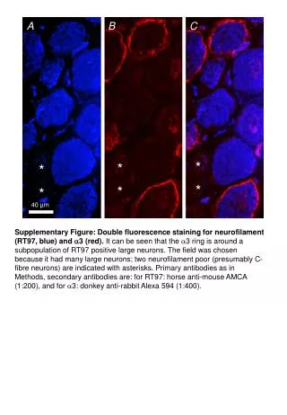
A B C * * * * * * Supplementary Figure:Double fluorescence staining for neurofilament (RT97, blue) and 3 (red). It can be seen that the 3 ring is around a subpopulation of RT97 positive large neurons. The field was chosen because it had many large neurons; two neurofilament poor (presumably C-fibre neurons) are indicated with asterisks. Primary antibodies as in Methods, secondary antibodies are: for RT97: horse anti-mouse AMCA (1:200), and for 3: donkey anti-rabbit Alexa 594 (1:400). 40 µm