Download

1 / 20
200 likes | 226 Vues
Learn about palatal tremors post-brainstem stroke, with MRI images and research on hypertrophic degeneration, acquired oscillations, and rhythmic muscle involvement. Explore the pacemaker theory, history, and treatment implications.

E N D
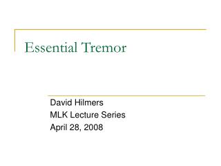
Palatal Tremor Shirley H. Wray, M.D., Ph.D. Professor of Neurology, Harvard Medical School Director, Unit for Neurovisual Disorders Massachusetts General Hospital
Neuroimaging Figure 1: Axial NECT scan shows a large pontine hemorrhage extending into the midbrain. This patient developed palatal tremor 2 years later.
Neuroimaging Figure 2: Axial T2WI in a patient who developed palatal tremor 6 months after a midbrain bleed from a cavernous malformation shows a small mixed signal intensity lesion in the dorsal midbrain tegmentum
Neuroimaging Figure 3: Axial T2WI (same case as Fig. 2) shows enlarged olives with striking hyperintensity characteristic for classic hypertrophic olivary degeneration
Palatal Tremor Continuous rhythmic movement of the soft palate Persists in sleep and coma Persists for life Asymptomatic Unilateral or midline
Palatal Tremor Frequently associated with time-locked synchronous oculo-pharyngo-laryngo-respiratory muscle involvement Latency 2 – 49 months mean 11 months post brainstem stroke Pathophysiology, hypertrophy of the inferior olivary nucleus
Acquired Pendular Oscillations* Symptomatic – oscillopsia Time locked with palatal myoclonus Suppressed in slow-wave sleep, present in REM sleep Can be present only on eye closure Senusoidal (pendular) wave form vertical/horizontal/elliptical Cold-caloric stimulation – no change Full eye movements *Frequently called ocular myoclonus
Figure 4. Direct current oculography showing vertical pendular ocular oscillations.
Figure 5. Schematic presentation of the main component of the ocular oscillations observed in the lateral form of OPM. They are reminiscient of ocular counter-rolling produced by head tilt about an anteroposterior axial lateral to the outer canthus of the eye (X).
The Inferior Olive – (IO) The observations support the view that the IO is the pacemaker Histological evidence of hypertrophic degeneration Normal IO cells have the capacity for spontaneous rhythmic discharges PET: IO hyperactive – increased glucose uptake
Oculopalatal TremorRhythmic Hyperactivity Release or disinhibition of different primitive rhythms in branchial muscles (Yakolev 1956) Rhythmicity on basis of denervation hypersensitivity of IO cells to transmitter (Matsuo & Ajax 1979) IO cells produce rhythmic synchronized discharges under special conditions (Llinas 1984, Llinas & Yarom 1986)
Figure 7. EEG, EMG and EOG recorded during natural nocturnal sleep in patient 1. EEG (F, frontal; C, central); EMI (submental); EMG 2 (right facial); EOG 1 (right eye); EOG 2 (left eye).
References Gallet J. Le Nystagmus du voile: Le syndrome myoclonique de la callotte protuberantielle thesis. Paris, 1927. Gullain G, Mollaret P. (1931) Deux cas de myoclonies synchrones et rythmees velo-pharyngo-laryngo-oculo diaphragmatiques. Le Probleme antomique et physio-pathologique de ce syndrome. Revue Neurologique, 2: 545-566.
Klein H. (1907) Zur Pathologic de kontinuierlichen rhythmischen Krampfe der schlingmuskulatur (2 Falle von Erweichungsherden im Kleinhirn). Neurologisches Centralblatt, 26: 245-254. Spencer HR. (1886) Pharyngeal and laryngeal “nystagmus”. Lancet 2: 702.