Intrauterine growth restriction
690 likes | 1.56k Vues
Intrauterine growth restriction. An evidence based approach. Introduction and background. IUGR remains a challenging problem for both the obstetrician and pediatrician.

Intrauterine growth restriction
E N D
Presentation Transcript
Intrauterine growth restriction An evidence based approach
Introduction and background • IUGR remains a challenging problem for both the obstetrician and pediatrician. • The ability to diagnose the disorder and understand its pathophysiology still outpaces the ability to prevent its complications..
Introduction and background • . Fetal growth restriction is the second leading cause of perinatal morbidity and mortality • . The incidence of intrauterine growth restriction (IUGR) is estimated to be approximately 5 percent in the general obstetric population
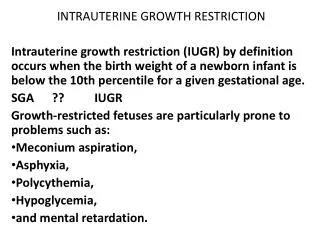
PERINATAL IMPLICATIONS IUGR causes a spectrum of perinatal complications, including • fetal deaths, prematurity, neonatal death, fetal compromise in labor, neonatal morbidity, induction of labor, and cesarean delivery. • Neonates with IUGR who survive the intrauterine experience are at increasedrisk for impaired neurodevelopment, and possibly type 2 diabetes and hypertension in adult life.
DEFINITIONS The term IUGR has changed from intrauterine growth retardation to the more current term, intrauterine growth restriction.. • IUGR:- a fetus is unable to grow to its genetically determined potential size to a degree that may affect the health of the fetus.
DEFINITIONS • SGA :-refer to a fetus that has failed to achieve a specific biometric or estimated weight threshold by a specific gestational age.. • The commonly used threshold is the tenth centile for abdominal circumference and estimated birthweight
DEFINITIONS • 50–70% of fetuses with a birthweight below tenth centile for gestational age are constitutionally small, and the lower the centile for defining SGA, the higher the likelihood of FGR • Not all fetuses who are SGA (<10th percentile) have IUGR, and not all fetuses who have IUGR are SGA. • For example, if a couple has had 3 term 4-kg babies and then has a 2.7-kg term baby, that infant is not SGA but may have IUGR.
Symmetric and Asymmetric IUGR • . Symmetric growth restriction implies a fetus whose entire body is proportionally small. • Asymmetric growth restriction implies a fetus who is undernourished and is directing most of its energy to maintaining growth of vital organs, such as the brain and heart, at the expense of the liver, muscle and fat. This type of growth restriction is usually the result of placental insufficiency.
Symmetric and Asymmetric IUGR • Investigators have differed on the importance of this differentiation in the diagnosis and management • But recently. The symmetrically grown infants who were SGA had outcomes very similar to the infants who were AGA. E-medicine May 2002
maternal causes Possible maternal causes of IUGR :- • Chronic hypertension • Pregnancy-associated hypertension • Cyanotic heart disease • Class F or higher diabetes • Hemoglobinopathies • Autoimmune disease • Protein-calorie malnutrition • Smoking • Substance abuse • Uterine malformations • Thrombophilia Severi, 2000
Fetal causes • Possible causes of IUGR related to the placenta and/or umbilical cord :- • Multiple gestation • Twin-to-twin transfusion • Abnormal cord insertion • Placental abnormality • Chronic abruption • Placenta previa • Cord anomalies • Identifying a cause may not be possible in as many as 40% of cases Severi, 2000
Diagnosis • Four important issues need to be considered : 1- most measurements require an accurate estimation of gestation as a prerequisite 2-most tests attempt to diagnose SGA fetuses rather than growth-restricted fetuses 3-most studies use a one-off measurement (size) to predict SGA while there is evidence that it is the trend (growth) that is of more value in predicting poor fetal outcome 4- no attention is made for important prognostic factors for SGA, such as maternal height, weight, ethnicity, parity and fetal gender
Diagnosis • It should also be noted that, although an individual test alone may not be predictive of SGA or FGR, a composite of abnormal results such as an ultrasonically small fetus with reduced liquor or abnormal uterine artery Doppler may indicate pathology.
Diagnosis . A distinction needs to be made between biometric tests (tests to measure size) and biophysical tests (tests to assess fetal wellbeing . the diagnosis of SGA would rely on biometric tests while abnormal biophysical tests are more indicative of FGR than SGA.
Diagnosis 1- Abdominal palpation • Grade C . Abdominal palpation has limited diagnostic accuracy to predict a SGA fetus. RCOG November 2002
Diagnosis 2- Fundal height • Grade B • Symphyseal fundal height (SFH) measurement has limited diagnostic accuracy to predict an SGA neonate. • Although early studies reported high sensitivity and specificity , a large study of 2941 women found the sensitivity and specificity to be 27% and 88%, respectively. • Serial measurements may improve sensitivity and specificity RCOG November 2002
Diagnosis 2- Fundal height • Grade B • A customised fundal height chart improves accuracy to predict a SGA fetus. • A customised SFH chart is adjusted for physiological variables such as maternal height, weight, parity and ethnic group. • Use of such charts was found to result in improvement in sensitivity . RCOG November 2002
Diagnosis3- Ultrasound biometry • GradeB A-Abdominal circumference and estimated fetal weight to diagnose SGA. • They are the most accurate diagnostic measurements to predict SGA. • In high-risk women, AC at less than the tenth centile has sensitivities of 72.9–94.5% and specificities of 50.6–83.8% in the prediction of fetuses with birthweight at less than the tenth centile. RCOG November 2002
Diagnosis 3- Ultrasound biometry • Grade B B-Customised ultrasound charts. • Customised ultrasound EFW charts that are adjusted for important independent physiological variables, such as maternal weight, maternal height, ethnic group and parity, have better sensitivities for identifying SGA fetuses , FGR, have lower false-positive rates and are predictive of poor perinatal events. RCOG November 2002
Diagnosis3- Ultrasound biometry • Grade B C- Growth velocity in addition to size. • Serial measurements of AC and EFW (growth velocities) are superior to single estimates of AC or EFW in the prediction of FGR and predicting poor perinatal outcome. • However, use of fetal growth alone to diagnose growth restriction (especially when the interval between the scan is less than two weeks) can lead to high numbers of false positives. RCOG November 2002
Diagnosis 3- Ultrasound biometry • Ratio measures, such as head to abdominal circumference (HC/AC) and femoral length to abdominal circumference (FL/AC) ratios are poorer than AC or EFW alone in predicting SGA. • A systematic review in the Cochrane Database of Systematic Reviews has shown that routine ultrasound after 24 weeks in low-risk pregnancy does not improve perinatal outcome Cochrane library Evidence level Ia
Diagnosis4- Biophysical tests • All biophysical tests, including amniotic fluid volume (AFV), Doppler, cardiotocography and biophysical scoring, are poor at diagnosing a small or growth-restricted fetus. RCOG November 2002
Diagnosis4- Biophysical tests • Grade B. • AFV has minimal value in diagnosing FGR • Despite the positive association between AFV and neonatal morphometry, the likelihood ratios remain low. • For amniotic fluid index (AFI), a positive test result has a likelihood ratio (LR) of 2.4 for predicting skinfold thickness below the tenth centile • Serial measurements of AFI have similarly disappointing results RCOG November 2002
Diagnosis 4- Biophysical tests • Grade A. • Uterine artery Doppler has limited use in predicting FGR. • A systematic review with meta-analysis published in 2000 found that uterine artery Doppler had limited accuracy in predicting FGR and perinatal death • Abnormal uterine artery suggest a maternal cause for the growth restriction where as normal uterine artery Doppler studies suggest that a fetal cause RCOG November 2002
management • At this point, the only reasonable goals in the treatment of IUGR are to deliver the most mature fetus in the best condition possible with minimal risk to the mother. Such a goal requires the use of antenatal testing in hope of identifying the fetus with IUGR before it becomes acidotic.
1- Assessment for chromosomal defects • When a small fetus is diagnosed, assess for risk of chromosomal defects. • Up to 19% of fetuses with an AC and EFW less than the fifth centile may have chromosomal defects • The risk is higher when growth restriction is associated with structural abnormalities, a normal liquor volume or a normal uterine or umbilical artery Doppler. • Therefore, all growth-restricted fetuses need an ultrasound anatomical survey as a minimum. It may also be appropriate to offer karyotyping.
2- Surveillance of the small fetus (umbilical artery Doppler ) • Grade A • Use umbilical artery Doppler as the primary surveillance tool. • A systematic review with meta-analysis has provided evidence that the use of umbilical artery Doppler to monitor high-risk fetuses reduces perinatal morbidity and mortality. • In addition, there was a significant reduction in the number of antenatal admissions and inductions of labour RCOG November 2002
umbilical artery Doppler • In normal pregnancy, there is a progressive increase in end-diastolic velocity due to growth and dilatation of the umbilical circulation. The resistance index therefore falls. • A resistance index > 0.72 is outside the normal limits from 26 weeks gestation onwards. • In some pregnancies with fetal growth restriction and/or preeclampsia, there is a reduction in the diastolic velocity and in severe cases, there is absent or reversed end diastolic velocity.
2- Surveillance of the small fetus (umbilical artery Doppler ) • . A study comparing fetal heart-rate monitoring, biophysical profile and umbilical artery Doppler found that only umbilical artery Doppler had value in predicting poor perinatal outcomes in SGA • Screening a low-risk or unselected population by umbilical artery Doppler,, is not recommended for screening.
2- Surveillance of the small fetus (umbilical artery Doppler ) • A variety of indices of umbilical arterial Doppler waveform, such as:- • resistance index, systolic/diastolic ratio, pulsatility index and diastolic average ratio, is used for predicting perinatal outcome. • resistance index had the best ability to predict abnormal outcomes • When an anomaly scan and umbilical artery Doppler are normal, the small fetus is likely to be a ‘normal small fetus • Evidence suggests that outpatient management of such fetuses is safe. RCOG November 2002 Evidence level II
2- Surveillance of the small fetus (umbilical artery Doppler ) • frequency of monitoring in SGA fetuses with normal Doppler need not generally be more than once every fortnight. RCOG November 2002 Evidence level II
2- Surveillanceof the small fetus (liquor volume ) • Grade B • Measure liquor volume using either AFI or pocket depth as both tests have similar diagnostic accuracy. • Abnormal liquor volume has been variously defined as single cord-free 1-cm, 2-cm, or an AFI below the fifth centile for the gestation or . 5 cm. RCOG November 2002
2- Surveillance of the small fetus (liquor volume ) • a systematic review with meta-analysis of 18 studies with over 10 000 patients found an antepartum AFI of . 5.0 cm was associated with an increased risk of an Apgar score of less than seven at five minutes RCOG November 2002Evidence level I and III
2- Surveillance of the small fetus (biophysical profile and cardiotocography ) • Grade A • The biophysical profile has not been shown to improve perinatal outcome but sufficient data do not exist to rule out its value. • However, there is evidence from uncontrolled observational studies that biophysical profile in high-risk women has good negative predictive value, i.e. fetal death is rare in women with a normal biophysical profile RCOG Nov. 2002 Evidence level Ia
2- Surveillance of the small fetus (biophysical profile and cardiotocography ) • the absence of benefit from randomised trials and since it is a time-consuming test, So it cannot be recommended for routine monitoring in low risk pregnancies or for primary surveillance in SGA • However, when primary surveillance with umbilical artery Doppler is found to be abnormal, biophysical profile is likely to be useful given its good negative predictive value in high-risk populations. • This recommendation is further supported by evidence that, in high-risk women, the biophysical profile was rarely abnormal when Doppler findings were normal. Evidence level Ib
2- Surveillance of the small fetus (biophysical profile and cardiotocography ) • Use of cardiotocography (CTG) antepartum to assess fetal condition is not associated with better perinatal outcome. • In fact, a systematic review of randomised trials showed that there was a trend towards increased mortality in the group receiving CTG compared with those who did not. Computer systems for interpretation of CTG have better accuracy than clinical experts in predicting umbilical acidosis and depressed Apgar scores. Evidence level Ib
Timing of delivery Delivery • There is wide variation in practice in the timing of delivery of growth restricted fetuses. RISK OF stillbirth Risk of prematurity
End diastolic flow is present (PED) • Grade C • When end diastolic flow is present (PED), delay delivery until at least 37 weeks, provided other surveillance findings are normal. • Absent or reversed end diastolic flow is associated with increased perinatal mortality and morbidity. The odds ratio for perinatal mortality with absent or revered end diastolic flow were 4.0 and 10.6, respectively, compared with when end diastolic flow was present. • The incidences of respiratory distress syndrome and necrotising enterocolitis were not increased with absent or revered end diastolic volume but there was an increase in cerebral haemorrhage, anaemia and hypoglycaemia. • Evidence level IIa
End diastolic flow is absent or reversed • Grade C When end diastolic flow is absent or reversed • admission, close surveillance and administration of steroids are required. • If other surveillance results (biophysical profile, venous Doppler) are abnormal, delivery is indicated. • If gestation is over 34 weeks, even if other results are normal, delivery may be considered.
If other surveillance results (biophysical profile,venous Doppler) are normal, and gestation is below34 weeks, The optimal surveillance strategy in those women is unclear. • Options include daily CTG/BPP and/or venous doppler deliver when • The CTG becomes pathological (decelerations with reduced variability), 2. The biophysical profile becomes abnormal 3.There is reversal of Doppler velocities in ductus venosus during atrial contraction 4.or there are umbilical vein pulsations. Under these circumstances, delivery is likely to be by caesarean section
If other surveillance results (biophysical profile,venous Doppler) are normal, and gestation is below34 weeks, • The interval between first occurrence of AED and an abnormal CTG/biophysical profile has ranged from 1–26 days depending on Gestational age, the presence of hypertension and venous Doppler abnormalities (notably pulsations in the umbilical vein)
Some forms of intervention • . Most prenatal interventions do not show any significant effects on perinatal outcome. • . Smoking cessation programmes, can be effective for a small minority of smokers in increasing birthweight but there are no data to suggest that this intervention improves perinatal outcome. • .
Some forms of intervention • Although a meta-analysis of 13 trials evaluated the use of aspirin in the prevention of growth restriction and found that it reduced the incidence of FGR, only a few studies have used aspirin in the treatment of FGR. These trials are small and have shown conflicting results. Further trials are needed to assess the value of aspirin in the treatment of FGR..
Some forms of intervention • A meta-analysis of randomized trials finds that a daily low dose of aspirin therapy among high-risk women significantly reduces the risk of perinatal death and preeclampsia also a significant reduction in the rate of spontaneous preterm births and an increase in birth weights • . The researchers did not find any evidence of harm from aspirin therapy in pregnant women. • They conclude that, aspirin therapy should be considered for pregnant women at high risk in prevention of IUGR. • ACOG May 31, 2003
Some forms of intervention There is not enough evidence to assess the value of • oxygen therapy, • nutrient therapy, • hospitalisation and bedrest, • betamimetics, • calcium channel blockers, • hormonal therapy • and plasma volume expansion in treating growth restriction. The Cochrane Library, Issue 3, 2003
Some forms of intervention • Grade A • Administer steroids if gestation is below 36 weeks. • Antenatal steroids significantly reduce the incidence of respiratory distress syndrome. RCOG November 2002