Download
1 / 123
1.23k likes | 1.34k Vues
Congenital cystic adenomatoid malformation. Findings: Multilocular low attn lesion in the RLL with thin enhancing septa ddx: Pulmonary abscess. Adrenal adenoma. Findings: Large right adrenal lesion Isointense to liver on in-phase scan

E N D
Congenital cysticadenomatoid malformation • Findings: • Multilocular low attn lesion in the RLL with thin enhancing septa • ddx: • Pulmonary abscess
Adrenal adenoma • Findings: • Large right adrenal lesion • Isointense to liver on in-phase scan • Hypointense to liver (signal loss) on out-of-phase scan • ddx: • NONE! • This is an Aunt Minnie!
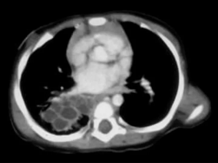
Pancreatic pseudocysts • Findings: • Multiple large pancreatic cystic lesions • ddx: • Von-Hippel Lindau • Cystic pancreatic neoplasm
Pulmonary hamartoma • Findings: • Solitary pulmonary fat-containing nodule in the RML • May exhibit “popcorn” calcification • ddx: • NONE! • This is an Aunt Minnie!
Adrenal macronodular hyperplasia • Findings: • Bilateral lobular enlargement of the adrenal glands. • ddx: • Cushing’s syndrome • Metastases • Infection • Tuberculosis • Histoplasmosis
Von-Hippel Lindau • Findings: • Numerous bilateral renal cyst • Solid enhancing right renal mass = RCC • ddx: • NONE! • This is an Aunt Minnie!
SVC obstruction • Findings: • Contrast-enhanced CT shows intense enhancement of the medial segment of the left lobe = “liver hot spot” sign • Collateral vessels seen anteriorly in the soft tissues • ddx: • NONE! • This is an Aunt Minnie!
Adrenal metastasis • Findings: • Large heterogeneous adrenal mass • Size alone makes it a surgical lesion • ddx: • Adenoma • Adenocarcinoma • Adrenal cortical carcinoma
Splenic infarct • Findings: • Large spleen containing a low attenuation geographic lesion extending from the hilus to the periphery • ddx: • NONE! • This is an Aunt Minnie!
Malignant mesothelioma • Findings: • Encasement and compression of left lung by enhancing soft tissue mass • Invasion of the posterolateral chest wall and mediastinum • ddx: • Metastases • Breast • Thymus
ADPKD • Findings: • Enlarged bilateral kidneys containing innumerable cysts • May also see hepatic cysts • Cysts complicated by hemorrhage or infection • NO increase risk of RCC • ddx: • NONE! • This is an Aunt Minnie!
Splenic hemangioma • Findings: • Low attenuation splenic lesion • Most common solid lesion of the spleen • Imaging and enhancement characteristics often unlike hepatic hemangiomas • ddx: • NONE! • This is an Aunt Minnie!
Nutmeg liver due toheart failure • Findings: • CT scan shows ascites and paradoxical enhancement of the liver during the arterial phase • ddx: • Hepatitis • Cirrhosis • Budd-Chiari
Appendix mucocele • Findings: • Smooth round filling defect at the cecal base • CT shows dilated tubular structure filled with low density material • ddx: • Mucinous adenocarcinoma of the appendix • ? Tubo-ovarian abscess
Calcified splenic cyst • Findings: • Large low density splenic lesion • Thick calcified wall • ddx: • Posttraumatic (false) • Epidermoid (true) • Chronic hydatid disease
Pneumacystis pneumonia • Findings: • Bilateral ground glass opacity emanating from the hila • Normal underlying parenchyma • ddx: • Hypersensitivity pneumonitis • Usual interstitial pneumonia • Cryptogenic organizing pneumonia • Hemorrhage
Focal nodular hyperplasia • Findings: • Intensely enhancing lesion in the hepatic dome in the arterial phase • Quick washout in the portal-venous phase • ddx: • Hypervascular tumor • Metastasis
Non-calcified splenic cyst • Findings: • Large homogeneous low density splenic lesion • No calcified wall • ddx: • Epidermoid cyst (true) • Pancreatic pseudocyst • Hydatid disease • Abscess
Right heart dysfunction • Findings: • Opacification of the hepatic veins during the arterial phase • Hepatic parenchyma unremarkable • ddx: • NONE! • This is an Aunt Minnie!
Cystic renal mass • Findings: • Low density renal mass with numerous thin septations • Partially exophytic, partially extending to the sinus • ddx: • Cystic RCC • Multilocular cystic nephroma • Abscess • Xanthogranulomatous pyelonephritis (focal)
Splenic arter aneurysm • Findings: • Dense round lesion medial to the splenic hilum • Same density as the aorta • Causes • Portal hypertension • Pancreatitis • FMD • ddx: • Hypervascular mass of stomach or pancreas
Thymic cyst • Findings: • Large cystic mass in the anterior mediastinum • High attenuation along the periphery = calcification or rim enhancement • ddx: • Cystic teratoma
Emphysematous cholecystitis • Findings: • Distended gallbladder with gas within the wall • Increased risk in pts. with DM • Surgical emergency • ddx: • NONE! • This is an Aunt Minnie!
Islet cell tumor • Findings: • Enhancing mass in the pancreatic tail = gastrinoma • Pancreatic cysts • Associations: • Z-E syndrome • VHL • MEN I • ddx: • Hypervascular metastasis