Download
1 / 36
360 likes | 558 Vues
Digitech Ophthalmologist. Product Overview. Digitech Ophthalmologist. What is Digitech Ophthalmologist ? Software tool for Ophthalmologist Its like a smart assistance who will carry out the routine tasks with ease freeing more time for the important work. Advantages

E N D
Digitech Ophthalmologist Product Overview
Digitech Ophthalmologist • What is Digitech Ophthalmologist ? • Software tool for Ophthalmologist • Its like a smart assistance who will carry out the routine tasks with ease freeing more time for the important work. • Advantages • This software operates on the images taken by Fundus Camera • Leaves more time for the doctor to actually focus on the important diagnostic part rather than menial work. • Presents information and statistics in such a way to help doctor take the best decision • Improves patient care at reduced prices as telemedicine is possible due to this innovation
Digitech Ophthalmologist Features: The major features are illustrated as below. • Patient Database Management • Distortion Correction • Panorama Image Construction • Cup to Disk Ration (CDR ) Analysis • Retina 3D Reconstruction • Blood Vessel Analysis • Glaucoma Detection • Retinopathy Each Feature is discussed at a greater length in the following slides.
Patient Database Management • Archiving and maintaining the medical data is one of the most important tasks • A tool for sophisticated archival and easy retrieval of the data is necessary when dealing with the large databases • Techlead Patient Database system avails doctors the following facilities • Store the database per patient basis for keeping track of history • Access the database for a particular clinical manifestation • Allow Multiple doctors to comment on the single case and all visible to the user • A portable format of database which can be shared among doctors with simpler means such as cell phones • Simple User Interface
Distortion Correction • Images are distorted due to the deviation of the light from direct Path. The distortion is induced by • EYE Lens • Camera Lens • Figure shows example of Camera Lens Distortion • Distortion Correction • Achieved by applying Polynomial Correction on the input image • Correction is applied on radial axis with the coefficients based on camera lens • Complete removal is impossible as eye lens characteristics of individual are not known
Panorama Construction • Complete retina surface is segmented in 7-9 virtual overlapping parts and individually photographed • Retina is photographed in such a way to compensate the distortion caused by the Eye lens and the curvature of retina. • Figure shows the method of photography of retina
Panorama Construction Cntd.. • Panorama Construction Features • Image Distortion is not 100% correctable making the alignment difficult • Manually Stitching Images is a tedious Job • Panorama construction utility performs this operation in no time • Testing • Tested on more than 100 datasets • Stitching time is < 40 seconds for 7 images (Each Image 9MPixel) • Time varies depending on the features in a image. Panorama Image is constructed from the 7 images 6 out of which are shown in the above slide taken at different positions (as shown with purple circles)
CDR Analysis • What is CDR ? • It’s the ratio between surface area occupied by Optic Cup to Surface area occupied by Optic Disc. • What’s its Clinical Significance • When Cup surface area increases, CDR increases. • This is indication of variation of intra ocular pressure and sometimes can indicate progress of glaucoma • Current Method • Manual marking – Ambiguous Optic Cup Contour Optic Disc Contour
CDR Analysis Cntd… • Accurate Extraction of the contours will result in the accurate prediction of clinical manifestation • How to increase the accuracy of detected contours • Active Contours or SNAKE algorithm • Like the snake , when put near the object, it fits the object contour with minimum error distance i.e. maximally touches the actual boundary • Example Video of Snake Operation
CDR Analysis Reports • CDR Analysis is used to monitor the intra ocular and dilation of Cup surface area • CDR is measured from the centre of the optic disk and traversing axially at a particular angle towards the disc contour. The distance where it cuts cup contour is marked as CUP while the distance at which it cuts the disc contour is marked as disc. • This procedure is repeated for 0 -360 degrees and is plot as a graph Centre point Cup Intercept Distance @ 0 Degree Angle 0 Degree Disc Intercept Distance @ 0 Degree
CDR Analysis Reports Figure shows the graph of CDR plot for two different dates for chronological studies
Blood Vessel Analysis • Tracing the blood vessels accurately is the backbone of the entire application • Blood vessels are used as the markers of information for panorama Image construction • AV Ratio gives vital information about Arterial narrowing and hence the systemic hypertension as well as Diabetics and optic atrophy • ALR or arterial reflex is a measure of thinning of the artery walls which in turn alarm about arteriosclerosis and atherosclerosis AV Ratio, Artery width to Vain Width
Development of the Blood Vessel Analysis Algorithm • Development of a robust algorithm for vessel tracing (Vessel Strokes algorithm ) • Development of Adaptive Vessel tracing algorithm (Accurate boundary detection for exact calculation of AV ratio and ALR are essential for Clinical manifestation) • Vessel classification and Tree formation (with some domain knowledge and fine tuning) • Fine tuning of the algorithm is required so that the best results are obtained for most of the images • The most important challenge still remains is to identify the algorithm which works for almost all the input images. Even the best algorithm needs to be fine tuned for the performance on a given dataset.
Robust Algorithm Development • Many algorithms were tried and tested for reliable extraction of the blood vessels from background in variety of images • Vessel Stroke algorithm performed the best in all kinds of input image conditions • Following figure shows example of vessel extraction
Adaptive Vessel Tracing Algorithm • Adaptive Vessel Tracing Algorithm • Drawing the centre line of the vessel • Drawing the boundaries of the vessel • Identifying the branch points • Identifying the crossover points • Identifying the end of the vessel • Accurately adjusting the boundaries as per local statistics • Identify and Join the broken vessels without addition of errors • Identify Blood vessel type: Artery / Vein • Build the Vessel Tree Data structure For each of the above parameters, the algorithm has been tested for more than 300 images at a time. This dataset consists of input images of various kinds.
Blood Vessel tracing Utility • The blood vessel tracing utility allows user to calculate the following • Traced Vessels • Vessel Classification- Artery and Veins Tree • Vessel Tree – Connected tree starts from Optic Disc • AV ratio at the selected region • ALR Ratio at the selected region • Following figure shows the snapshot of the utility
3D Retina Reconstruction • 3D analysis is a powerful tool for in biomedical applications and already popular in radiology • Retina Can be viewed as a 2D image as well as a 3D structure • 3D visualization is possible if the stereo view of the Retina is available • Once the stereo imaging data is in hand getting the 3D output is a two step job • Generation of 3D data from 2D Images using stereo algorithms • Visualizing the 3D data with some viewer
Generation of 3D Data from 2D images • Stereo imaging takes 2 images with a lateral shift, preferably from two different cameras. • The two images are processed to identify the disparity between the matching structures to calculate the depth of the feature. • Figure below shows one such example
Features of Techlead Stereo Algorithm • Robust algorithm even works for images taken by single camera. Ideal if the input is provided from a standard stereo imaging setup with 2 cameras • Automatic Y shift correction • Automatic Disparity range calculation • Automatic Cropping of the Area of Interest • Automatic correction of rotation • Auto Identification of left and right images • Facility to fine tune parameters (defaults are stored) • View the output in a interactive 3D viewer
Glaucoma Detection • What is Glaucoma? • Glaucoma is a disease which can result in loss of vision. • It’s early detection and treatment is of prime importance. • Physiological Appearance: Fan-like pattern near Optic Cup Extended Optic Cup
Glaucoma Detection (Contd..) • Evaluation of Nerve Fiber layer(NFL) is essential for early detection of Glaucoma. • Physiological Appearance of NFL: • Methods of Glaucoma Detection • Median Radial Transform • Fourier Series Descriptors • Hough Transform Striated Parabolic Arcs
Glaucoma Detection Results Glaucoma Detection NFL Detection
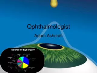
Retinopathy • Retinopathy refers to a group of diseases which can cause severe vision loss or blindness. • This retinal damage can be a result of ruptured blood vessels, swelling in optic nerve etc. • Digital Retinopathy identifies these symptoms by the abnormal coloring as shown below:
Retinopathy (Contd..) Retinopathy Algorithm Detected Abnormal Patch
Retinopathy : Screening • Screening • If the input image is normal or abnormal • User can train the software with • Selecting and deleting the reference Images. • Accepting / rejecting the outcome of the software • Adjusting thresholds • Internal Algorithm will tune itself to give best results
Retinopathy: Training • Salient Features: • Training – the doctor can train this software to classify what are normal and abnormal images. Input Algorithm Output Training Feedback
Retinopathy (Contd..) • Salient Features: • Intelligent Disease Locator • Uses biological domain knowledge to extract the diseased areas • Train the algorithm to separate a particular pattern of data • Use this knowledge for future reference with systematic classification
Retinopathy (Contd..) • Retinopathy Disease Tracker Tool: • Analyze large data • View Patient History • Patient’s Response • Graphical representation
Retinopathy (Contd..) • Retinopathy Disease Tracker Tool(Contd..) • Shows statistics along with data sets. • Finds similar patient history. • Software will present analysis and hypothesis to doctors to be accepted or rejected. • Content based image retrieval used to identify similar cases. • Doctor can share the data of a particular patient case with another in a standard format like DICOM including all the analysis and expert comments.
Retinopathy (Contd..) • Multiple Examinations • When one patient comes more than one time • The current analysis is checked with history and the results are shown under multiple examinations Newly identified area Marked with Blue Circle