Download
1 / 31
310 likes | 312 Vues
This article discusses the case of a young postpartum woman who was transported to the emergency room in full cardiopulmonary arrest due to venous air embolism. The article explains the causes, symptoms, diagnosis, and management of venous air embolism.

E N D
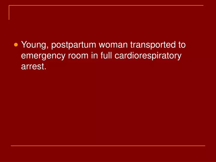
Young, postpartum woman transported to emergency room in full cardiorespiratory arrest.
Cause of Death • Venous air embolism. • Due to vaginal insufflation. • Other significant conditions: • Postpartum subinvolution of uterus.
Embolism: an obstruction in a blood vessel due to a blood clot or foreign material which occludes the vessel after traveling through the blood stream.
Types of emboli • Thromboemboli • Fat • Air • Tumor • Bullets
Venous Air Emboli • Air/gas introduced into systemic venous circulation. • Many cases are probably subclinical and resolve without significant consequences.
Required Conditions • Direct communication between the source of air and vasculature. • Pressure gradient favoring the passage of gas into the venous circulation.
Important Factors • Amount of air introduced into the venous circulation. • Rate of accumulation of air in venous circulation. • Position of patient.
Quantity of Gas • Small amounts of air are broken up and reabsorbed in the capillary beds. • 100 to 200 ml have been reported in the literature as potentially fatal. • > 5 ml/kg. • Significant symptoms have been reported with as little as 20 ml of air (length unprimed IV tubing). • 2-3 ml into cerebral circulation can be fatal. • 0.5 ml into the LAD can cause ventricular fibrillation.
Rate of Air Infusion • Rapid infusion of air overwhelms the capacity of the capillaries and leads to right ventricular strain with outflow obstruction, increased pulmonary artery pressure and ultimately cardiovascular failure.
Frequency • True incidence is unknown. • Radiologic literature reports 0.13% during insertion and removal of central venous catheters with optimal technique. • 1/47 to 1/3000 of CVC. • 10-50% of high risk neurosurgical procedures.
Morbidity and Mortality • Dependent on multiple factors: • Amount and rate of air infusion. • Other associated conditions.
Causes • Surgical procedures: • Neurosurgical procedures especially with patient in sitting position or with open dural sinuses. • Trauma surgery with compromised large vessels. • Tumors with high vascularity.
Causes • OB-GYN procedures. • Central venous catheters: • Central lines. pulmonary catheters, hemodialysis catheters, Hickman lines, etc. • Mechanical insufflation: • Orogenital sexual activity. • Inadvertent injection of air with IV contrast or other radiologic studies.
Diagnosis • Must have a high suspicion as clinical symptoms can be non-specific or mimic other conditions.
History • Recent surgical procedures. • Blunt or penetrating trauma of head, neck, chest or abdomen • Central venous catheterization. • Peripostpartum oral genital sexual activity.
Clinical Symptoms • Will be dependent on the amount of air and rate of effusion. • Position of patient (sitting versus recumbent). • Paradoxical arterial embolism.
Presentation • Symptoms include shortness of breath, cough, nausea, chest pain, agitation (impending doom) and disorientation. • Cardiovascular findings: • dysrhythmias, hypotension, pulmonary hypertension, myocardial ischemia, increased central venous pressure, “mill wheel murmur”, nonspecific ECG findings (ST segment and T wave changes), shock.
Presentation • Pulmonary findings: • Abnormal breath sounds (rales and wheezing), tachypnea, hemoptysis, cyanosis, decreased arterial oxygen saturation, elevated CO2, increased pulmonary artery pressures, pulmonary edema.
Presentation • Neurologic: • Altered mental status, seizures, focal deficits, loss of consciousness, coma. • Ophthalmologic: • Air bubbles in retinal vessels • Skin: • Crepitus over superficial vessels.
Differential Diagnosis • Extensive and can include coronary artery syndrome, aortic stenosis, atrial fibrillation, bronchospasm, COPD, CHF, aortic dissection, CVA, metabolic disorders, pulmonary emboli and shock from multiple causes.
Diagnostic Studies • Laboratory: • No specific tests. • Blood gas. • Specific tests may be useful for assessing organ damage.
Diagnostic Studies • Imaging studies: • Transesophageal echocardiography. • High sensitivity for detecting air in right ventricle and outflow tract. • Precordial Doppler ultrasound. • Chest x-ray. • CT scan.
Diagnostic Studies • ECG • Non-specific findings (right ventricular strain, increased heart rate, ST depression). • Pulse oximetry • Central venous catheter aspiration.
Management • Transport patient in left lateral decubitus position. • Stop any procedure immediately. • Can attempt to remove air via catheter. • Provide hemodynamic support. • 100% oxygen/intubation. • Maintain volume. • Vasopressors and mechanical ventilation. • Consider hyperbaric oxygen therapy if neurologic symptoms or cardiac instability