Download
1 / 60
1.69k likes | 8.4k Vues
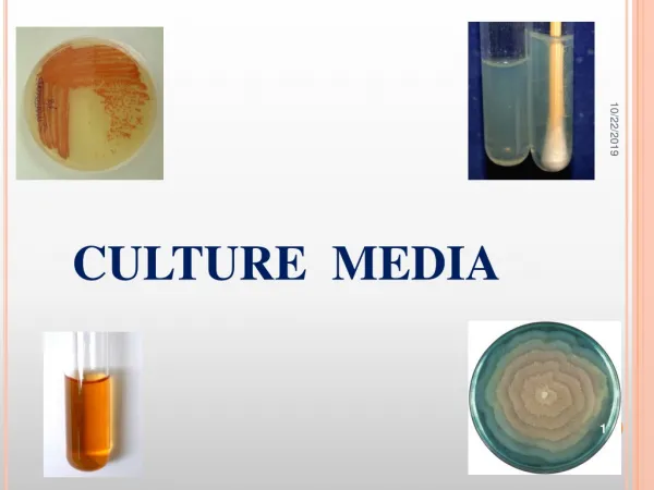
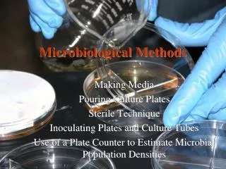
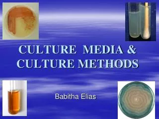
CULTURE MEDIA & CULTURE METHODS. Babitha Elias. Bacteria have to be grown (cultured) for them to be identified. By appropriate procedures they have to be grown separately (isolated) on culture media and obtained as pure for study. History

E N D
CULTURE MEDIA & CULTURE METHODS Babitha Elias
Bacteria have to be grown (cultured) for them to be identified. By appropriate procedures they have to be grown separately (isolated) on culture media and obtained as pure for study. History The original media used by Louis Pasteur – urine or meat broth Liquid medium – diffuse growth Solid medium – discrete colonies.
Colony– macroscopically visible collection of millions of bacteria originating from a single bacterial cell. Cooked cut potato by Robert Koch – earliest solid medium Gelatin – not satisfactory - liquefy at 24oC
Agar Frau Hesse Used for preparing solid medium Obtained from seaweeds. No nutritive value Not affected by the growth of the bacteria. Melts at 98oC & sets at 42oC 2% agar is employed in solid medium
Types of culture media Based on their consistency a) solid medium b) liquid medium c) semi solid medium Based on the constituents/ ingredients a) simple medium b) complex medium c) synthetic or defined medium d) Special media
Special media Enriched media Enrichment media Selective media Indicator media Differential media Sugar media Transport media Media for biochemical reactions Based on Oxygen requirement - Aerobic media - Anaerobic media
Solid media – contains 2% agar Colony morphology, pigmentation, hemolysis can be appreciated. Eg: Nutrient agar, Blood agar Liquid media – no agar. For inoculum preparation, Blood culture, for the isolation of pathogens from a mixture. Eg: Nutrient broth Semi solid medium – 0.5% agar. Eg: Motility medium
Simple media / basal media - Eg: NB, NA - NB consists of peptone, meat extract, NaCl, - NB + 2% agar = Nutrient agar
Complex media Media other than basal media. They have added ingredients. Provide special nutrients Synthetic or defined media Media prepared from pure chemical substances and its exact composition is known Eg: peptone water – 1% peptone + 0.5% NaCl in water
Enriched media Substances like blood, serum, egg are added to the basal medium. Used to grow bacteria that are exacting in their nutritional needs. Eg: Blood agar, Chocolate agar
Chocolate agar Blood agar
Enrichment media • Liquid media used to isolate pathogens from a mixed culture. • Media is incorporated with inhibitory substances to suppress the unwanted organism. • Eg: • Selenite F Broth – for the isolation of Salmonella, Shigella • Alkaline Peptone Water – for Vibrio cholerae
Selective media The inhibitory substance is added to a solid media. Eg: Mac Conkey’s medium for gram negative bacteria TCBS – for V.cholerae LJ medium – M.tuberculosis Wilson and Blair medium – S.typhi Potassium tellurite medium – Diphtheria bacilli
Mac Conkey’s medium TCBS
Potassium Tellurite media LJ media
Indicator media • These media contain an indicator which changes its colour when a bacterium grows in them. • Eg: • Blood agar • Mac Conkey’s medium • Christensen’s urease medium
Differential media • A media which has substances incorporated in it enabling it to distinguish between bacteria. • Eg: Mac Conkey’s medium • Peptone • Lactose • Agar • Neutral red • Taurocholate • Distinguish between lactose fermenters & non lactose fermenters.
Lactose fermenters – Pink colonies Non lactose fermenters – colourless colonies
Sugar media Media containing any fermentable substance. Eg: glucose, arabinose, lactose, starch etc. Media consists of 1% of the sugar in peptone water. Contain a small tube (Durham’s tube) for the detection of gas by the bacteria.
Transport media • Media used for transporting the samples. • Delicate organisms may not survive the time taken for transporting the specimen without a transport media. • Eg: • Stuart’s medium – non nutrient soft agar gel containing a reducing agent • Buffered glycerol saline – enteric bacilli
Anaerobic media These media are used to grow anaerobic organisms. Eg: Robertson’s cooked meat medium, Thioglycolate medium.
BIOCHEMICAL TEST & REACTIONS They provide additional information for the identification of the bacterium. The tests include: Oxidase test Triple sugar iron agar (TSI) Indole test Citrate utilization Urease test
OXIDASE TEST Detects the presence of an enzyme “oxidase” produced by certain bacteria which will reduce the dye – tetramethyl-p-phenylene diamine dihydrochloride. Positive test is indicated by the development of a purplecolour. Oxidase positive – Pseudomonas, Vibrio, Neisseriae Oxidase negative – Salmonella, Shigella
TRIPLE SUGAR IRON AGAR (TSI) It is a composite media used to study different properties of a bacterium – sugar fermentation, gas production and H2S production. In addition to peptone, yeast extract & agar, it contains 3 sugars – Glucose, Lactose, Sucrose. The Iron salt – Ferric citrate indicates H2S production. Phenol red is the indicator. It is an orange red medium with a slant and a butt. pH of the medium – 7.4
TSI REACTIONS: Yellow – Acid Pink - Alkaline Yellow slant / Yellow butt (A/A) – Lactose fermenters. Pink slant / Yellow butt (K/A) – Non lactose fermenters. Pink slant / no colour change (K/K) – Non fermenters Black colour – H2S production. Gas bubbles or crack in the medium – gas production. LF – E.coli, Klebsiella NLF – Salmonella, Shigella H2S - Proteus
INDOLE TEST Used to detect indole production by the organism. They produce indole from tryptophan present in peptone water. After overnight incubation, a few drops of indole reagent (Kovac’s reagent) is added. Positive test is indicated by a pink ring. Positive indole test – pink ring Negative indole test - yellow ring Indole positive – E.coli Indole negative – Klebsiella, Salmonella.
CITRATE UTILIZATION • Done in Simmon’s Citrate medium. • To detect the ability of certain bacteria to utilize citrate as the sole source of carbon. • Contains Sodium citrate and bromothymol blue as the indicator. • If citrate is utilized, alkali is produced which turns the medium to blue. • Citrate positive – blue colour • Citrate negative – green colour • Positive – Klebsiella • Negative – E.coli
UREASE TEST • Done in Christensen’s urease medium. • This test is used to detect organisms that produce urease. • Urease produced by the organisms split urea into ammonia and CO2. • Urease positive – pink colour • Urease negative – yellow colour • Positive – Proteus, Klebsiella • Negative – E.coli, Salmonella
CULTURE METHODS • Culture methods employed depend on the purpose for which they are intended. • The indications for culture are: • To isolate bacteria in pure cultures. • To demonstrate their properties. • To obtain sufficient growth for the preparation of antigens and for other tests. • For bacteriophage & bacteriocin susceptibility. • To determine sensitivity to antibiotics. • To estimate viable counts. • Maintain stock cultures.
Culture methods include: Streak culture Lawn culture Stroke culture Stab culture Pour plate method Liquid culture Anaerobic culture methods
STREAK CULTURE Used for the isolation of bacteria in pure culture from clinical specimens. Platinum wire or Nichrome wire is used. One loopful of the specimen is transferred onto the surface of a well dried plate. Spread over a small area at the periphery. The inoculum is then distributed thinly over the plate by streaking it with a loop in a series of parallel lines in different segments of the plate. On incubation, separated colonies are obtained over the last series of streaks.
LAWN CULTURE Provides a uniform surface growth of the bacterium. Uses For bacteriophage typing. Antibiotic sensitivity testing. In the preparation of bacterial antigens and vaccines. Lawn cultures are prepared by flooding the surface of the plate with a liquid suspension of the bacterium.
STROKE CULTURE Stroke culture is made in tubes containing agar slope / slant. Uses Provide a pure growth of bacterium for slide agglutination and other diagnostic tests.
STAB CULTURE Prepared by puncturing a suitable medium – gelatin or glucose agar with a long, straight, charged wire. Uses Demonstration of gelatin liquefaction. Oxygen requirements of the bacterium under study. Maintenance of stoke cultures.
Gelatin liquefaction Oxidation – Fermentation medium
POUR PLATE CULTURE Agar medium is melted (15 ml) and cooled to 45oC. 1 ml of the inoculum is added to the molten agar. Mix well and pour to a sterile petri dish. Allow it to set. Incubate at 37oC, colonies will be distributed throughout the depth of the medium. Uses Gives an estimate of the viable bacterial count in a suspension. For the quantitative urine cultures.
LIQUID CULTURES Liquid cultures are inoculated by touching with a charged loop or by adding the inoculum with pipettes or syringes. Uses Blood culture Sterility tests Continuous culture methods Disadvantage It does not provide a pure culture from mixed inocula.